. 2010 Jun;38(11):3743-59.
doi: 10.1093/nar/gkq054. Epub 2010 Feb 15.

Abundance of type I toxin-antitoxin systems in bacteria: searches for new candidates and discovery of novel families

Affiliations

Abundance of type I toxin-antitoxin systems in bacteria: searches for new candidates and discovery of novel families

Elizabeth M Fozo et al. Nucleic Acids Res. 2010 Jun.

Abstract

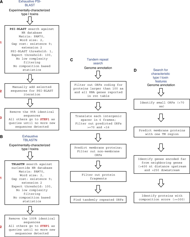
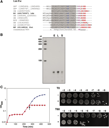
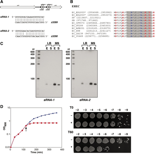
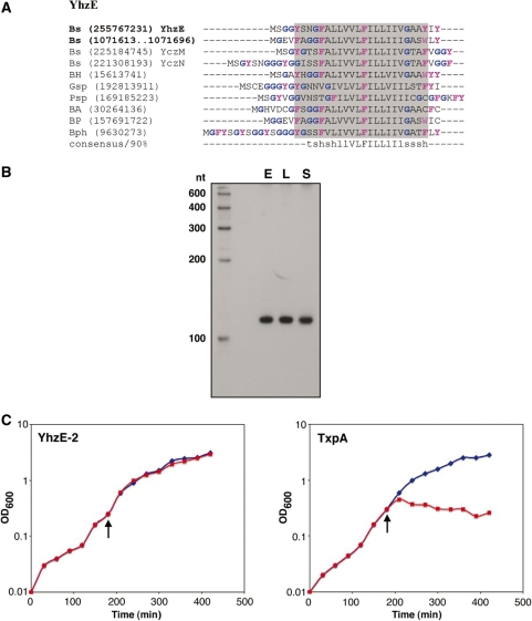
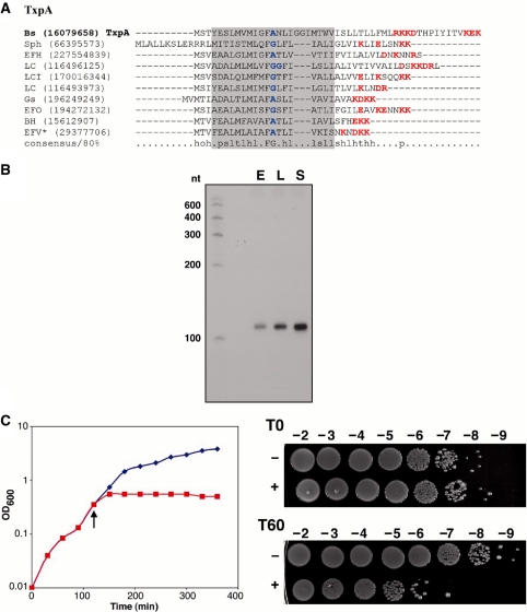
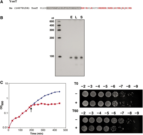
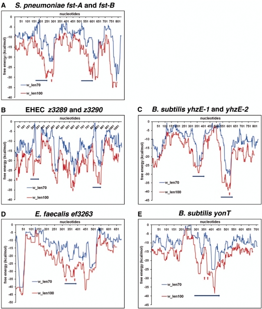
Small, hydrophobic proteins whose synthesis is repressed by small RNAs (sRNAs), denoted type I toxin-antitoxin modules, were first discovered on plasmids where they regulate plasmid stability, but were subsequently found on a few bacterial chromosomes. We used exhaustive PSI-BLAST and TBLASTN searches across 774 bacterial genomes to identify homologs of known type I toxins. These searches substantially expanded the collection of predicted type I toxins, revealed homology of the Ldr and Fst toxins, and suggested that type I toxin-antitoxin loci are not spread by horizontal gene transfer. To discover novel type I toxin-antitoxin systems, we developed a set of search parameters based on characteristics of known loci including the presence of tandem repeats and clusters of charged and bulky amino acids at the C-termini of short proteins containing predicted transmembrane regions. We detected sRNAs for three predicted toxins from enterohemorrhagic Escherichia coli and Bacillus subtilis, and showed that two of the respective proteins indeed are toxic when overexpressed. We also demonstrated that the local free-energy minima of RNA folding can be used to detect the positions of the sRNA genes. Our results suggest that type I toxin-antitoxin modules are much more widely distributed among bacteria than previously appreciated.


Figures

Figure 1.
Figure 1.
Computational approaches used to identify and predict type I toxins. (A) Exhaustive PSI-BLAST to identify homologs of known toxins not among previously annotated protein sequences. (B) Exhaustive TBLASTN search to identify homologs of known type I toxins not among previous annotated ORFs. (C) Tandem repeat search to identify new type I toxins encoded in the same intergenic region. (D) Search for new type I toxins based upon characteristics of known toxins.
Figure 2.
Figure 2.
(A) Multiple alignment of selected representatives of the Ldr/Fst family. Proteins studied in this work are denoted by asterisk, experimentally characterized type I toxins are shown in bold; the predicted transmembrane region is shaded; conserved small amino acids are colored blue; the conserved tryptophan is colored magenta; charged amino acids (RKDE) are colored red. The consensus was built using CONSENSUS program (http://coot.embl.de/Alignment//consensus.html) for a larger set of Ldr/Fst proteins (see Supplementary Figure S1A). The sequences are denoted by both the abbreviated species name and the GI number or the coordinates in the corresponding genome in parentheses. Species abbreviations: SP, Streptococcus pneumoniae R6; CB, Clostridium bolteae; EC, Escherichia coli K-12 substr. MG1655; SB, Shigella boydii CDC 3083-94; SE, Salmonella enterica arizonae z4z23; ECO, Escherichia coli O127:H6 str. E2348/69; SA, Staphylococcus aureus ssp. aureus Mu50; LG, Lactobacillus gasseri MV-22; EF, Enterococcus faecalis plasmid pAD1. (B) Northern blot showing expression of an sRNA antisense to S. pneumoniae fst-B-homolog. Total RNA (10 µg) isolated from S. pneumoniae R6 cells grown to OD600 ≈ 0.3 (E), OD600 ≈ 1.0 (L) and OD600 ≈ 1.5 (S) in BHI medium was loaded in each lane. (C) Overproduction of the S. pneumoniae Fst-B homolog in E. coli. MG1655 harboring pAZ3-fst-B was grown in LB medium to OD600 ≈ 0.3. The culture was split (indicated by the arrow); half was left untreated (blue) while arabinose (0.2% final concentration) was added to the other half (red). Cell dilutions were plated 0 (T0) and 60 (T60) min following arabinose induction.
Figure 3.
Figure 3.
(A) Genomic arrangement of EHEC Z3289 and Z3290. The ORFs are indicated by the black regions of the leftward arrows and the regions of complementarity are indicated by the white boxes. The sequences capable of base pairing are shown below the gene arrangement. (B) Multiple alignment of selected representatives of EHEC family. Most designations are the same as in the Figure 2A. The predicted transmembrane regions is shaded (predicted for E. coli O157:H7 EDL933 proteins and extended for other sequences): small amino acids are colored blue; charged amino acids (RKDE) are colored red. Species abbreviations (strains are also indicated for E. coli species): EC, E. coli; SB, Shigella boydii CDC 3083-94; SF, Shigella flexneri 2a str. 2457T. (C) Expression of the antitoxin RNAs for Z3289 (sRNA-1) and Z3290 (sRNA-2). Total RNA (10 µg) isolated from E. coli O157:H7 EDL933 cells grown to OD600 ≈ 0.4 (E) and OD600 ≈ 5.0 (overnight, S) in LB medium and from cells grown to OD600 ≈ 0.4 (E) and OD600 ≈ 2.2 (overnight, S) in M9 media supplemented with 0.2% glucose was loaded in each lane. (D) Overproduction of Z3290 in MG1655. MG1655 harboring pAZ3-z3290 was grown in LB medium to OD600 ≈ 0.3. The culture was split (indicated by the arrow); half was left untreated (blue) while arabinose (0.2% final concentration) was added to the other half (red). Cell dilutions were plated 0 (T0) and 60 (T60) min following arabinose induction.
Figure 4.
Figure 4.
(A) Multiple alignment of selected representatives of YhzE family. Most designations are the same as in the Figure 2A. The consensus was built using CONSENSUS program for a larger set of YhzE family proteins (see Supplementary Figure S1C). The predicted transmembrane regions is shaded (predicted for B. subtilis YhzE protein and extended for other sequences); small amino acids are colored blue; aromatic residues are colored magenta. Species abbreviations: Bs, B. subtilis str. 168; Bph, Bacillus phage SPBc2; Gsp, Geobacillus sp. G11MC16; BH, B. halodurans C-125; BA, B. anthracis str. Ames; Psp, Paenibacillus sp. JDR-2; BP, B. pumilus SAFR-032. (B) Expression of an sRNA antisense to yhzE-2. Total RNA (10 µg) isolated from B. subtilis PY79 cells grown to OD600 ≈ 0.3 (E), OD600 ≈ 2.0 (L) and OD600 ≈ 3.5 (S) in LB medium was loaded in each lane. (C) Overproduction of YhzE-2 and TxpA in B. subtilis PY79. YhzE-2 (graph on the left) or TxpA (right) under the control of the Plac promoter was integrated into the amyE locus of PY79. The cultures were grown in LB medium to OD600 ≈ 0.3. The cultures were split; (indicated by the arrow); half was left untreated (blue) while IPTG (1 mM final concentration) was added to the other half (red).
Figure 5.
Figure 5.
(A) Multiple alignment of selected representatives of the TxpA family. The consensus was built using CONSENSUS program for a larger set of the TxpA family proteins (see Supplementary Figure S1D). Most designations are the same as in the Figure 2A. The predicted transmembrane regions is shaded (predicted for Enterococcus faecalis V583 protein and extended for other sequences). Species abbreviations: Bs, B. subtilis sub. subtilis str. 168; Sph, Staphylococcus phage 42E; EFH, E. faecalis HH22; LC, Lactobacillus casei ATCC 334; LCI, Leuconostoc citreum KM20; LC, Lactobacillus casei ATCC 334; Gs, Geobacillus sp. G11MC16; EFO, E. faecalis OG1RF; BH, B. halodurans C-125; EFV, E. faecalis V583. (B) Northern blot showing expression of an sRNA antisense to EF3263 in E. faecalis OG1RF. Total RNA (10 µg) isolated from E. faecalis OG1RF cells grown to OD600 ≈ 0.3 (E), OD600 ≈ 1.0 (L) and OD600 ≈ 1.5 (S) in BHI medium was loaded in each lane. (C) Overproduction of EF3263 in E. coli. MG1655 harboring pAZ3-ef3263 was grown in LB medium to OD600 ≈ 0.3. The culture was split (indicated by the arrow); half was left untreated (blue) while arabinose (0.2% final concentration) was added to the other half (red). Cell dilutions were plated 0 (T0) and 60 (T60) min following arabinose induction.
Figure 6.
Figure 6.
(A) Amino acid sequence of yonT gene product of Bacillus subtilis ssp. subtilis str. 168 (Bs). Charged amino acids (EKR) are colored red and the predicted transmembrane regions is shaded. (B) Expression of an sRNA antisense to yonT. Total RNA (10 µg) isolated from B. subtilis ssp. subtilis str. 168 cells grown to OD600 ≈ 0.3 (E), OD600 ≈ 2.0 (L) and OD600 ≈ 3.5 (S) in LB medium was loaded in each lane. A smaller band of ∼80 nt can be seen upon overexposure as the cells enter the stationary phase of growth. (C) Overproduction of YonT in E. coli. MG1655 harboring pAZ3-yonT was grown in LB medium to OD600 ≈ 0.3. The culture was split (indicated by the arrow); half was left untreated (blue) while arabinose (0.2% final concentration) was added to the other half (red). Cell dilutions were plated 0 (T0) and 60 (T60) min following arabinose induction.
Figure 7.
Figure 7.
Prediction of antitoxin sRNAs using free-energy profiles for RNA local secondary structures. Free-energy profiles for RNA local secondary structures along nucleotide sequences in experimentally tested RNA antitoxin systems in S. pneumoniae (A), E. coli (B), B. subtilis (C and E) and E. faecalis (D). The lengths of the sliding window used for free-energy estimations (70 and 100 nt) corresponded to common lengths of the previously described sRNA antitoxins. Blue arrows show location of predicted ORFs. Red arrows show the positions of the oligonucleotides used to detect the antisense sRNAs. Other local free-energy minima correspond to annotated terminators or unrelated short ORFs. x-axis: nucleotide positions; y-axis: free energy of RNA folding.

References

    1. Fozo EM, Hemm MR, Storz G. Small toxic proteins and the antisense RNAs that repress them. Microbiol. Mol. Biol. Rev. 2008;72:579–589. - PMC - PubMed
    1. Gerdes K, Wagner E.GH. RNA antitoxins. Curr. Opin. Microbiol. 2007;10:117–124. - PubMed
    1. Makarova KS, Wolf YI, Koonin EV. Comprehensive comparative-genomic analysis of type 2 toxin-antitoxin systems and related mobile stress response systems in prokaryotes. Biol. Direct. 2009;4:19. - PMC - PubMed
    1. Yamaguchi Y, Inouye M. mRNA interferases, sequence-specific endoribonucleases from the toxin-antitoxin systems. Prog. Mol. Biol. Transl. Sci. 2009;85:467–500. - PubMed
    1. Gerdes K, Christensen SK, Løbner-Olesen A. Prokaryotic toxin-antitoxin stress response loci. Nat. Rev. Microbiol. 2005;3:371–382. - PubMed

Publication types

MeSH terms