. 2010 Mar;133(Pt 3):797-807.
doi: 10.1093/brain/awq015. Epub 2010 Feb 15.

Mechanism of neurodegeneration of neurons with mitochondrial DNA mutations

Affiliations

Mechanism of neurodegeneration of neurons with mitochondrial DNA mutations

Andrey Y Abramov et al. Brain. 2010 Mar.

Abstract

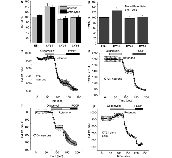
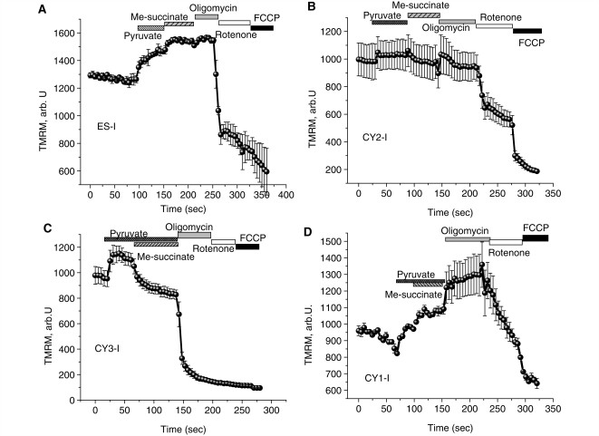
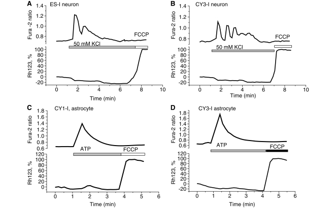
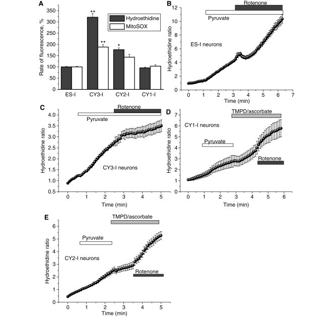
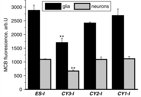
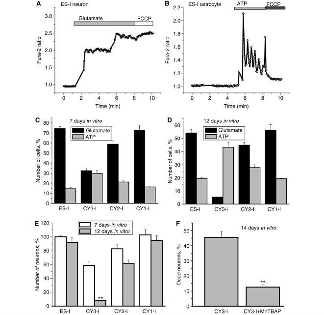
Mutations of mitochondrial DNA are associated with a wide spectrum of disorders, primarily affecting the central nervous system and muscle function. The specific consequences of mitochondrial DNA mutations for neuronal pathophysiology are not understood. In order to explore the impact of mitochondrial mutations on neuronal biochemistry and physiology, we have used fluorescence imaging techniques to examine changes in mitochondrial function in neurons differentiated from mouse embryonic stem-cell cybrids containing mitochondrial DNA polymorphic variants or mutations. Surprisingly, in neurons carrying a severe mutation in respiratory complex I (<10% residual complex I activity) the mitochondrial membrane potential was significantly increased, but collapsed in response to oligomycin, suggesting that the mitochondrial membrane potential was maintained by the F(1)F(o) ATPase operating in 'reverse' mode. In cells with a mutation in complex IV causing approximately 40% residual complex IV activity, the mitochondrial membrane potential was not significantly different from controls. The rate of generation of mitochondrial reactive oxygen species, measured using hydroethidium and signals from the mitochondrially targeted hydroethidine, was increased in neurons with both the complex I and complex IV mutations. Glutathione was depleted, suggesting significant oxidative stress in neurons with a complex I deficiency, but not in those with a complex IV defect. In the neurons with complex I deficiency but not the complex IV defect, neuronal death was increased and was attenuated by reactive oxygen species scavengers. Thus, in neurons with a severe mutation of complex I, the maintenance of a high potential by F(1)F(o) ATPase activity combined with an impaired respiratory chain causes oxidative stress which promotes cell death.


Figures

Figure 1
Figure 1
Characteristics of mitochondrial membrane potential (Δψm) in cells with mitochondrial mutations. (A–B) Neurons and astrocytes with severe mutation in complex I (CY3-I) showed a significant increase (P < 0.001) in Δψm compared with control cells. The mitochondrial potential in cells with a mutation in complex IV (CY2-I) showed no significant difference from control. Non-differentiated CY3-I cells also exhibited a 26% increase (P < 0.05) in tetramethylrhodamine methylester (TMRM) fluorescence (i.e. an increased Δψm) compared with controls. In control and CY2-I neurons (C–D), oligomycin did not affect Δψm; rotenone induced a partial depolarization; FCCP induced complete depolarization. In CY3-I neurons (E), oligomycin caused a mitochondrial depolarization. In the CY3-I stem cells (F) the Δψm was not maintained by reverse electron flow since oligomycin did not cause mitochondrial depolarization. *P < 0.05; **P < 0.001.
Figure 2
Figure 2
Effect of mitochondrial substrates on mechanism of maintenance of Δψm in cells with mitochondrial mutations. Application of pyruvate (5 mM) or methyl succinate (5 mM) to neurons increased Δψm, but increased substrate provision did not prevent the oligomycin induced mitochondrial depolarization in CY3-I neurons (C). In the presence of pyruvate, methyl-succinate induced further hyperpolarization of mitochondria in ES-I and CY1-I neurons (A, B, D), but a small mitochondrial depolarisation in CY3-I cells. TMRM = tetramethylrhodamine methylester.
Figure 3
Figure 3
Calcium homeostasis. Simultaneous measurements of [Ca2+]c and ΔΨm were made from neurons (A–B) and astrocytes (C–D) in mixed culture co-loaded with fura-2 and rhodamine 123. Traces are shown from single cells in each case. Mitochondrial mutations did not induce pathological changes in their mitochondrial responses to [Ca2+]c signals under these conditions.
Figure 4
Figure 4
Mitochondrial and cytosolic reactive oxygen species production in cells with mitochondrial mutations. CY3-I neurons displayed a significantly higher basal rate of increase in mitochondrially targeted hydroethidine (Mitosox) and hydroethidine ratio, demonstrating a higher basal production of intra-mitochondrial and extra-mitochondrial reactive oxygen species compared with control (A). Histogram demonstrating percentage values of the rate of mitochondrially targeted hydroethidine or hydroethidine ratio compared with 100% for control neurons. (B–E) show increase of ΔΨm by mitochondrial substrates (pyruvate and TMPD/ascorbate) or inhibition of complex 1 with rotenone demonstrated the dependence of reactive oxygen species production on ΔΨm. *P < 0.05; **P < 0.001.
Figure 5
Figure 5
Glutathione in both neurons and astrocytes. MCB was used to assess astrocyte and neuronal glutathione concentration. Mean intensities of MCB-glutathione adduct fluorescence (arb.U) at a steady state are presented. In CY3-I glutathione concentration was decreased compared with other cell lines in both astrocytes and neurons.
Figure 6
Figure 6
Cell death in neurons and astrocytes. The number of functional neurons and astrocytes in a co-culture was estimated by counting cells that show [Ca2+]c signals in response to physiological stimuli, which give characteristic and distinct responses in either neurons (glutamate, 50 μM) (A) or astrocytes (ATP, 100 μM) (B). With increasing time in culture, there was a progressive increase in cell death, seen predominantly in neurons, resulting in a change in the proportion between cell types (C–E). The antioxidant MnTBAP significantly protected CY3-I cells against the progressive increase in cell death; the number of dead neurons in this experiment was estimated as percentage of propidium iodide-positive neurons in the culture (F). *P < 0.05.

Similar articles

Cited by

References

    1. Abramov AY, Duchen MR. Mechanisms underlying the loss of mitochondrial membrane potential in glutamate excitotoxicity. Biochim Biophys Acta. 2008;1777:953–64. - PubMed
    1. Abramov AY, Scorziello A, Duchen MR. Three distinct mechanisms generate oxygen free radicals in neurons and contribute to cell death during anoxia and reoxygenation. J Neurosci. 2007;27:1129–38. - PMC - PubMed
    1. Bender A, Krishnan KJ, Morris CM, Taylor GA, Reeve AK, Perry RH, et al. High levels of mitochondrial DNA deletions in substantia nigra neurons in aging and Parkinson disease. Nat Genet. 2006;38:515–7. - PubMed
    1. Bender A, Schwarzkopf RM, McMillan A, Krishnan KJ, Rieder G, Neumann M, et al. Dopaminergic midbrain neurons are the prime target for mitochondrial DNA deletions. J Neurol. 2008;255:1231–5. - PubMed
    1. Betts J, Jaros E, Perry RH, Schaefer AM, Taylor RW, Abdel-All Z, et al. Molecular neuropathology of MELAS: level of heteroplasmy in individual neurones and evidence of extensive vascular involvement. Neuropathol Appl Neurobiol. 2006;32:359–73. - PubMed

Publication types

MeSH terms