. 2010 Apr 23;285(17):12765-77.
doi: 10.1074/jbc.M109.045161. Epub 2010 Feb 15.

A structural determinant that renders G alpha(i) sensitive to activation by GIV/girdin is required to promote cell migration

Affiliations

A structural determinant that renders G alpha(i) sensitive to activation by GIV/girdin is required to promote cell migration

Mikel Garcia-Marcos et al. J Biol Chem. .

Abstract

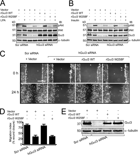
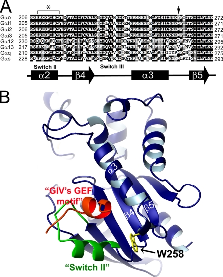
Although several non-receptor activators of heterotrimeric G proteins have been identified, the structural features of G proteins that determine their interaction with such activators and the subsequent biological effects are poorly understood. Here we investigated the structural determinants in G alpha(i3) necessary for its regulation by GIV/girdin, a guanine-nucleotide exchange factor (GEF) that activates G alpha(i) subunits. Using G protein activity and in vitro pulldown assays we demonstrate that G alpha(i3) is a better substrate for GIV than the highly homologous G alpha(o). We identified Trp-258 in the G alpha(i) subunit as a novel structural determinant for GIV binding by comparing GIV binding to G alpha(i3)/G alpha(o) chimeras. Mutation of Trp-258 to the corresponding Phe in G alpha(o) decreased GIV binding in vitro and in cultured cells but did not perturb interaction with other G alpha-binding partners, i.e. G betagamma, AGS3 (a guanine nucleotide dissociation inhibitor), GAIP/RGS19 (a GTPase-activating protein), and LPAR1 (a G protein-coupled receptor). Activation of G alpha(i3) by GIV was also dramatically reduced when Trp-258 was replaced with Tyr, Leu, Ser, His, Asp, or Ala, highlighting that Trp is required for maximal activation. Moreover, when mutant G alpha(i3) W258F was expressed in HeLa cells they failed to undergo cell migration and to enhance Akt signaling after growth factor or G protein-coupled receptor stimulation. Thus activation of G alpha(i3) by GIV is essential for biological functions associated with G alpha(i3) activation. In conclusion, we have discovered a novel structural determinant on G alpha(i) that plays a key role in defining the selectivity and efficiency of the GEF activity of GIV on G alpha(i) and that represents an attractive target site for designing small molecules to disrupt the G alpha(i)-GIV interface for therapeutic purposes.


Figures

FIGURE 1.
FIGURE 1.
GIV is a bona fide GEF for Gαi3. A, His-GIV-CT (aa 1623–1870) and His-GIV-CTs (aa 1660–1870) are equally efficient in increasing the steady-state GTPase activity of Gαi3. The steady-state GTPase activity of purified His-Gαi3 (50 nm) was determined in the presence of the indicated amounts (0, 0.1, 0.25, 0.5, 1, and 2 μm) of purified His-GIV-CT (aa 1623–1870, closed circles) or His-GIV-CTs (aa 1660–1870, open circles) by quantification of the amount of [γ-32P]GTP (0.5 μm, ∼50 cpm/fmol) hydrolyzed in 10 min. Data are expressed as % of GTP hydrolyzed by the G protein alone (0 μm His-GIV-CT or His-GIV-CTs). Results are shown as mean ± S.E. of n = 3 independent experiments. B, His-GIV-CTs does not affect the rate of GTP hydrolysis by Gαi3. Single-turnover GTPase assays for Gαi3 (50 nm) were performed as described under “Experimental Procedures” in the presence of His-GIV-CTs (2 μm, open circles), GST-GAIP (1 μm, “x”), or buffer (closed circles). GST-GAIP increases the rate of GTP hydrolysis by Gαi3, whereas His-GIV-CTs has no effect. One representative experiment of four is shown (C) His-GIV-CTs increases GTPγS binding to Gαi3. Nucleotide binding activity of purified His-Gαi3 (50 nm) was determined in the presence of the indicated amounts (0, 0.1, 0.5, 1, and 2 μm) of purified wild-type His-GIV-CTs (closed circles) or His-GIV-CTs F1685A mutant (open circles) by quantification of the amount of [35S]GTPγS (0.5 μm, ∼50 cpm/fmol) bound in 15 min. Data are expressed as % of GTPγS bound by the G protein alone in the absence of His-GIV-CTs. His-GIV-CTs increases GTPγS binding up to 2.2-fold over basal binding, whereas His-GIV-CTs F1685A has no significant effect. Results are shown as mean ± S.E. of n = 3 independent experiments.
FIGURE 2.
FIGURE 2.
GIV binds to and activates Gαi3 more efficiently than Gαo. A, GST-Gαi3·GDP (lane 1) binds ∼15- to 20-fold more endogenous GIV than GST-Gαo·GDP (lane 3). No binding of either Gαi3 or Gαo is detected in the presence of AlF4 (lanes 2 and 4). COS7 cell lysates (∼250 μg of protein) were incubated with ∼10 μg of purified GST-Gαi3 (lanes 1 and 2) or GST-Gαo (lanes 3 and 4) pre-loaded with GDP (lanes 1 and 3) or GDP·AlF4 (lanes 2 and 4) immobilized on glutathione beads. After extensive washing, bound proteins were separated by SDS-PAGE and analyzed by immunoblotting (IB) for GIV. Equal loading of GST proteins was confirmed by Ponceau S staining (lower panel). B, GST-Gαi3·GDP (lane 1) binds ∼10-fold more His-GIV-CT than GST-Gαo·GDP (lane 3). His-GIV-CT binding to both GST-Gαi3 and GST-Gαo is dramatically reduced in the presence of AlF4 (lanes 2 and 4). ∼3 μg of purified His-GIV-CT (aa 1623–1870) was incubated with ∼10 μg of purified GST-Gαi3 (lanes 1 and 2) or GST-Gαo (lanes 3 and 4) pre-loaded with GDP (lanes 1 and 3) or GDP·AlF4 (lanes 2 and 4) immobilized on glutathione beads. After extensive washing, bound proteins were separated by SDS-PAGE and analyzed by immunoblotting (IB) for His. Equal loading of GST proteins was confirmed by Ponceau S-staining (lower panel). C, His-GIV-CTs increases steady-state GTP hydrolysis of His-Gαi3 at least 3-fold more than His-Gαo at all concentrations tested. Results are shown as mean ± S.E. of nine independent experiments. The steady-state GTPase activity of purified His-Gαi3 (closed circles, 50 nm) or His-Gαo (open circles, 50 nm) was determined in the presence of the indicated amounts (0, 0.1, 0.5, and 2 μm) of purified His-GIV-CTs (aa 1660–1870) by quantification of the amount of [γ-32P]GTP (0.5 μm, ∼50 cpm/fmol) hydrolyzed in 10 min. Data is expressed as % of GTP hydrolyzed by the G protein alone in the absence of His-GIV-CTs.
FIGURE 3.
FIGURE 3.
Amino acids 178–270 of Gαi3 are responsible for the preferential binding of GIV to Gαi3versuso. A, schematic showing the Gαi3/o chimeras (1–4) used in B. In B: Upper panel, GDP-loaded GST-Gαi3/o chimera 1 (lane 6) and chimera 3 (lane 10) bind as much full-length GIV as GST-Gαi3 (lane 4), whereas GST-Gαi3/o chimera 2 (lane 8) and chimera 4 (lane 12) show dramatically reduced GIV binding. Binding of GIV to GDP·AlF4-loaded G proteins is absent in all cases (lanes 3, 5, 7, 9, 11, and 13). COS7 cell lysates were incubated with purified GST (lanes 2 and 3), GST-Gαi3 (lanes 4 and 5) or the indicated GST-Gαi3/o chimeras (lanes 6–13) pre-loaded with GDP (lanes 2, 4, 6, 8, 10, and 12) or GDP·AlF4 (lanes 3, 5, 7, 9, 11, and 13) immobilized on glutathione beads and analyzed as in Fig. 2A. A higher exposure of the same immunoblot (middle panel) shows the weak binding observed for GST-Gαi3/o chimera 2 (lane 8) and 4 (lane 12). Equal loading of GST proteins was confirmed by Ponceau S staining (lower panel).
FIGURE 4.
FIGURE 4.
Trp-258 of Gαi3 is responsible for GIV binding and activation of Gαi3. A, sequence alignment of Gαo, Gαi1, Gαi2, and Gαi3 indicating the Gαi3 mutants studied. Rat Gαo, Gαi1, Gαi2, and Gαi3 sequences corresponding to Gαi3 aa 178–270 were obtained form the NCBI data base and aligned using ClustalW. Conserved identical residues are in black; similar residues are shaded in gray. The secondary structure elements (α = α-helix, β = β-sheet) indicated below the alignment are named according to their crystal structures (29, 33). Residues conserved among Gαi1, Gαi2, and Gαi3 but different in Gαo within the 178–270 region were mutated in GST-Gαi3 to the corresponding residue in Gαo (indicated above with arrows). B, mutations W258F/T260I and W258F, in the α3/β5 loop, impair endogenous GIV binding to GDP-loaded GST-Gαi3, whereas mutations G217D, L232Q, A235H/E239T/M240T, and K248M do not affect binding (upper panels). GIV binding to GDP·AlF4-loaded G proteins is virtually absent in all cases (lower panels). COS7 cell lysates were incubated with ∼5 μg of purified GST-Gαi3 or the indicated GST-Gαi3 mutants pre-loaded with GDP (upper panels) or GDP·AlF4 (lower panels) immobilized on glutathione beads and analyzed as in Fig. 2A. Equal loading of GST proteins was confirmed by Ponceau S staining. C, binding of GST-GIV-CT to His-Gαi3 W258F (lane 6) is reduced (∼80%) compared with wt His-Gαi3 (lane 3). ∼3 μg of purified wt His-Gαi3 (lanes 1–3) or His-Gαi3 W258F (lanes 4–6) pre-loaded with GDP were incubated with ∼9 μg of GST (lanes 2 and 5) or GST-GIV-CT (aa 1623–1870, lanes 3 and 6) immobilized on glutathione beads. Bound proteins were analyzed by immunoblotting (IB) for His. Equal loading of GST proteins was confirmed by Ponceau S staining (lower panel). D, mutation of Trp-258 to Phe reduces (∼60–70%) activation of His-Gαi3 by His-GIV-CTs at all concentrations tested. The steady-state GTPase activity of purified His-Gαi3 (closed circles, 50 nm) or His-Gαi3 W258F (open circles, 50 nm) was determined as in Fig. 2C. Results are shown as mean ± S.E. of n = 9 (Gαi3) or n = 5 (Gαi3 W258F) independent experiments. E, mutation of Phe-259 to Trp increases (∼2-fold) activation of His-Gαo by His-GIV-CTs at all concentrations tested. The steady-state GTPase activity of purified His-Gαo (closed circles, 50 nm) or His-Gαo F259W (open circles, 50 nm) was determined as in Fig. 2C. Results are shown as mean ± S.E. of three independent experiments.
FIGURE 5.
FIGURE 5.
i3 activation by GIV is reduced when Trp-258 is replaced by Tyr, Leu, Ser, His, Asp, or Ala. A, all the Gαi3 Trp-258 mutants investigated show reduced activation by His-GIV-CT ranging from a ∼60% (W258Y) to ∼95% (W258D) reduction. Trp-258 of His-Gαi3 was mutated to amino acids of different nature, i.e. tyrosine (aromatic), leucine (aliphatic), serine (polar), histidine (basic), aspartate (acidic), and alanine (small). The steady-state GTPase activity of purified His-Gαi3 (closed circles, 50 nm) or the indicated His-Gαi3 mutants (open circles, 50 nm) was determined as described in Fig. 1C. Results are shown as mean ± S.E. of 4–9 independent experiments. B, binding of GST-GIV CT to His-Gαi3 mutants W258F (lane 3), W258A (lane 9), and W258D (lane 12) is reduced compared to wt His-Gαi3 (lane 6). ∼3 μg of purified His-Gαi3 (lanes 4–6) or the indicated His-Gαi3 mutants (W258F: lanes 1–3, W258A: lanes 7–9, W258D: lanes 10–12) pre-loaded with GDP were incubated with ∼9 μg of GST (lanes 2, 5, 8, and 11) or GST-GIV-CT (aa 1623–1870, lanes 3, 6, 9, and 12) immobilized on glutathione beads. Bound proteins were analyzed by immunoblotting (IB) for His. Equal loading of GST proteins was confirmed by Ponceau S staining (lower panel).
FIGURE 6.
FIGURE 6.
Mutations at Trp-258 do not affect the trypsin sensitivity of Gαi3 after activation by AlF4.i3 wt and all the Gα mutants tested are digested when pre-loaded with GDP, and all adopt the trypsin-resistant conformation after incubation with GDP·AlF4. His-Gαi3 and the indicated His-Gαi3 mutants or His-Gαo (0.5 mg/ml) were incubated in the presence of GDP or GDP·AlF4 and treated or not with trypsin as described under “Experimental Procedures.” A Coomassie Blue-stained gel of a representative experiment is shown. The arrowhead denotes the position of the non-trypsinized full-length proteins loaded, and the asterisk arrowhead denotes the trypsin-resistant form of the active, GDP·AlF4-loaded His-Gα subunit.
FIGURE 7.
FIGURE 7.
Mutation of Trp-258 to Asp, Ala, or Phe impairs the interaction between Gαi3 and endogenous GIV in cultured cells. Upper panel: co-immunoprecipitation of GIV, Gβγ, and AGS3 with FLAG-tagged Gαi3 mutants W258D (lane 2) or W258A (lane 3) is dramatically reduced compared with wt Gαi3 (lane 4). By contrast, with Gαi3 W258F (lane 5) only co-immunoprecipitation of GIV, but not Gβγ and AGS3 is reduced compared to controls. COS7 cells were transfected with empty vector (lane 1) or plasmids encoding FLAG-tagged Gαi3 W258D (lane 2), Gαi3 W258A (lane 3), wt Gαi3 (lane 4), or Gαi3 W258F (lane 5). 48 h after transfection cells were harvested, and lysates were used for immunoprecipitation (IP) with anti-FLAG IgG (∼2 μg) as described under “Experimental Procedures.” IP was followed by immunoblotting (IB) for FLAG (Gαi3), GIV, AGS3, and Gβ (pan-Gβ). Equal IgG loading was confirmed by Ponceau S staining. Lower panel: aliquots of the lysates (10%) were analyzed by immunoblotting (IB) to confirm the equal loading of Gαi3, GIV, AGS3, and Gβ and the expression of Gαi3-FLAG constructs in the different transfected samples.
FIGURE 8.
FIGURE 8.
i3 W258F binds GAIP and LPAR1 as efficiently as wt Gαi3. A, mutation of Trp-258 to Phe does not affect Gαi3 interaction with GAIP. GDP·AlF4-preloaded Gαi3 W258F (lane 6, upper panel) binds GAIP to the same extent as wt Gαi3 (lane 3, upper panel). Neither of the GDP-loaded G proteins binds GAIP (lanes 3 and 6, lower panel). ∼2 μg of purified wt His-Gαi3 (lanes 1–3) or His-Gαi3 W258F (lanes 4–6) pre-loaded with GDP·AlF4 (upper panel) or GDP (lower panel) were incubated with ∼5 μg of GST (lanes 2 and 5) or GST-GAIP (lanes 3 and 6) immobilized on glutathione beads. Bound proteins were analyzed by immunoblotting (IB) for His. 10% of the input His-Gα proteins were loaded in lanes 1 and 4. Equal loading of GST proteins was confirmed by Ponceau S staining. In B: Upper panel, FLAG-tagged LPAR1 (lanes 4–6) co-immunoprecipitates with both exogenously expressed wt Gαi3 (lane 5) and Gαi3 W258F mutant (lane 6). Endogenous Gαi3 (lane 4) is not detected in FLAG-LPAR1 immunoprecipitates, and no Gαi3 is present in FLAG-immunoprecipitates from controls (lanes 1–3). 48 h after transfection cells were harvested and lysates used for immunoprecipitation (IP) with anti-FLAG IgG (∼2 μg) as described under “Experimental Procedures.” IP was followed by immunoblotting (IB) for FLAG (LPAR1) and Gαi3. Equal IgG loading was confirmed by Ponceau S staining. Lower panel, aliquots of cell lysates (∼5%) were analyzed for expression of FLAG (LPAR1), Gαi3, and α-tubulin by immunoblotting (IB) to confirm the expression of the analyzed proteins.
FIGURE 9.
FIGURE 9.
i3 W258F fails to rescue Akt activation and cell migration defects observed upon depletion of endogenous Gαi3. A and B, HeLa cells treated with scrambled (Scr) or hGαi3 siRNA oligonucleotides and the indicated DNA plasmids (empty vector, rGαi3 WT, or rGαi3 W258F) were serum-starved for 6 h and stimulated with 10 μm LPA (A) or 100 nm insulin (B) for 5 min. Cell lysates were analyzed by immunoblotting (IB) for total (tAkt) and S473 phospho-Akt (pAkt), Gαi3, and α-tubulin. Depletion of endogenous Gαi3 reduces LPA-stimulated (A) and insulin-stimulated (B) Akt activation by ∼70%, which is restored upon transfection of wt rGαi3 but not rGαi3 W258F. C, in controls (Scr siRNA), HeLa cells cover the majority of the experimental wound area after 24 h, whereas in Gαi3-depleted cells wound closure is greatly impaired. The ability to migrate and close the wound area at 24 h is restored by transfection of rGαi3 wt but not rGαi3 W258F. HeLa cell monolayers treated with the indicated siRNA oligonucleotides, and DNA plasmids were scratch-wounded and examined by light microscopy immediately (0 h) or 24 h after wounding. Scale bar = 500 μm. D, bar graph showing quantification of the wound area covered by cells in C. The area covered by cells was determined by calculating the difference between the wound area at 0 and 24 h expressed as percent of the wound area at 0 h. Results are shown as mean ± S.D. of 8–12 randomly chosen fields from three independent experiments. E, cell lysates from cells treated as in C were immunoblotted to assess the efficiency of siRNA depletion of Gαi3 (∼95%) and the expression of rGαi3 WT or rGαi3 W258F.
FIGURE 10.
FIGURE 10.
Sequence comparison and three-dimensional view of the newly identified structural determinant in Gαi3 required for its activation by GIV. A, representative members of different Gα subfamilies were aligned to compare the sequence corresponding to the newly identified structural determinant in Gαi3. Alignment of rat Gαo, Gαi1, Gαi2, Gαi3, Gα12, Gα13, Gαq, and Gαs sequences obtained form the NCBI data base was performed using ClustalW. Conserved identical residues are in black; similar residues are shaded in gray. The secondary structure elements (α = α-helix, β = β-sheet) corresponding to these sequences are named according to previously reported crystal structures (29, 33) and are indicated below the alignment. The arrow denotes the position corresponding to the Trp-258 of Gαi3, and the asterisk denotes the previously identified binding site for GIV (11). B, three-dimensional view of the GEF motif of GIV bound to Gαi3 is shown to depict the relative location of the newly identified structural determinant (Trp-258 in the α3/β5 loop), which is positioned C-terminal to the GEF motif. The homology model of GDP·Gαi3 in complex with the GEF motif of GIV generated as described previously (11) using the structure of the synthetic peptide KB-752 bound to Gαi1 (PDB: 1Y3A) as a template (18). The “Ras-like” domain of Gαi3 is shown in blue, the “switch II” region in green, and Trp-258 in yellow. The GEF motif of GIV is shown in red.

Similar articles

Cited by

References

    1. De Vries L., Zheng B., Fischer T., Elenko E., Farquhar M. G. (2000) Annu. Rev. Pharmacol. Toxicol. 40, 235–271 - PubMed
    1. Gilman A. G. (1987) Annu. Rev. Biochem. 56, 615–649 - PubMed
    1. Ross E. M., Wilkie T. M. (2000) Annu. Rev. Biochem. 69, 795–827 - PubMed
    1. Oldham W. M., Hamm H. E. (2008) Nat. Rev. Mol. Cell Biol. 9, 60–71 - PubMed
    1. Overington J. P., Al-Lazikani B., Hopkins A. L. (2006) Nat. Rev. Drug Discov. 5, 993–996 - PubMed

Publication types

MeSH terms