

Effects of sex, race, and puberty on cortical bone and the functional muscle bone unit in children, adolescents, and young adults
- PMID: 20157194
- PMCID: PMC2853999
- DOI: 10.1210/jc.2009-1913
Effects of sex, race, and puberty on cortical bone and the functional muscle bone unit in children, adolescents, and young adults
Abstract
Context: Sex and race differences in bone development are associated with differences in growth, maturation, and body composition.
Objective: The aim of the study was to determine the independent effects of sex, race, and puberty on cortical bone development and muscle-bone relations in children and young adults.
Design and participants: We conducted a cross-sectional study of 665 healthy participants (310 male, 306 black) ages 5-35 yr.
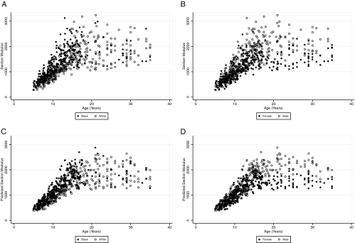
Outcomes: Tibia peripheral quantitative computed tomography measures were made of cortical bone mineral content (BMC) and bone mineral density (BMD), periosteal (Peri) and endosteal circumferences, section modulus (Zp), and muscle area. Regression models were adjusted for tibia length, age, race, sex, and Tanner stage.
Results: All cortical measures were greater in blacks than whites (all P < or = 0.001) in Tanner stages 1-4; however, differences in BMC, Peri, and Zp were negligible in Tanner stage 5 (all interactions, P < 0.01). Cortical BMC, Peri, and Zp were lower in females than males in all Tanner stages (all P < 0.001), and the sex differences in Peri and Zp were greater in Tanner stage 5 (interaction, P < 0.02). Cortical BMD was greater (P < 0.0001) and endosteal circumference was lower (P < 0.01) in Tanner 3-5 females, compared with males. Adjustment for muscle area attenuated but did not eliminate sex and race differences in cortical dimensions. Associations between muscle and bone outcomes did not differ according to sex or race.
Conclusion: Sex and race were associated with maturation-specific differences in cortical BMD and dimensions that were not fully explained by differences in bone length or muscle. No race or sex differences in the functional muscle bone unit were identified.
Figures

References
-
- Duan Y, Beck TJ, Wang XF, Seeman E 2003 Structural and biomechanical basis of sexual dimorphism in femoral neck fragility has its origins in growth and aging. J Bone Miner Res 18:1766–1774 - PubMed
-
- Seeman E 2002 Pathogenesis of bone fragility in women and men. Lancet 359:1841–1850 - PubMed
-
- Baron JA, Barrett J, Malenka D, Fisher E, Kniffin W, Bubolz T, Tosteson T 1994 Racial differences in fracture risk. Epidemiology 5:42–47 - PubMed
-
- Rauch F, Schoenau E 2001 The developing bone: slave or master of its cells and molecules? Pediatr Res 50:309–314 - PubMed
Publication types
MeSH terms
Grants and funding
- R01 DK064966/DK/NIDDK NIH HHS/United States
- R01-DK60030/DK/NIDDK NIH HHS/United States
- R01-HD040714/HD/NICHD NIH HHS/United States
- K24-DK076808/DK/NIDDK NIH HHS/United States
- UL1-RR024134/RR/NCRR NIH HHS/United States
- T32 DA023356/DA/NIDA NIH HHS/United States
- T32 DK060455/DK/NIDDK NIH HHS/United States
- K24 DK076808/DK/NIDDK NIH HHS/United States
- R01 HD040714/HD/NICHD NIH HHS/United States
- R01 DK060030/DK/NIDDK NIH HHS/United States
- UL1 RR024134/RR/NCRR NIH HHS/United States
- R01-DK064966/DK/NIDDK NIH HHS/United States
