

Microstructural diffusion changes are independent of macrostructural volume loss in moderate to severe Alzheimer's disease
- PMID: 20157252
- PMCID: PMC2889147
- DOI: 10.3233/JAD-2010-1295
Microstructural diffusion changes are independent of macrostructural volume loss in moderate to severe Alzheimer's disease
Abstract
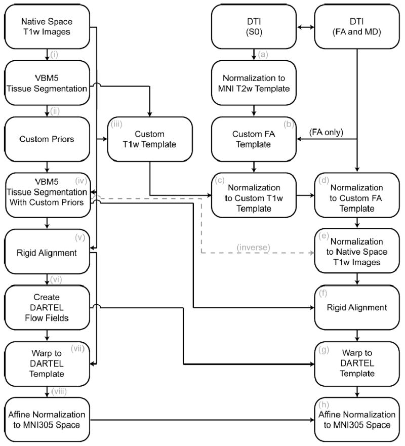
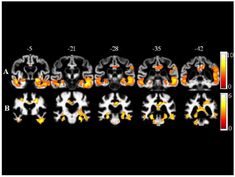
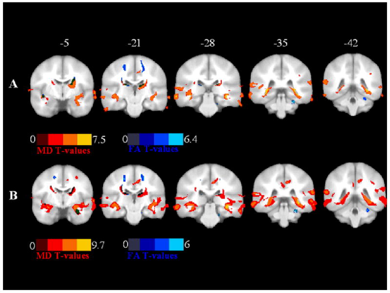
Although it is established that Alzheimer's disease (AD) leads to cerebral macrostructural atrophy, microstructural diffusion changes have also been observed, but it is not yet known whether these changes offer unique information about the disease pathology. Thus, a multi-modal imaging study was conducted to determine the independent contribution of each modality in moderate to severe AD. Seventeen patients with moderate-severe AD and 13 healthy volunteers underwent diffusion-weighted and T1-weighted MR scanning. Images were processed to obtain measures of macrostructural atrophy (gray and white matter volumes) and microstructural damage (fractional anisotropy and mean diffusivity). Microstructural diffusion changes independent of macrostructural loss were investigated using an ANCOVA where macrostructural maps were used as voxel-wise covariates. The reverse ANCOVA model was also assessed, where macrostructural loss was the dependent variable and microstructural diffusion tensor imaging maps were the imaging covariates. Diffusion differences between patients and controls were observed after controlling for volumetric differences in medial temporal, retrosplenial regions, anterior commissure, corona radiata, internal capsule, thalamus, corticopontine tracts, cerebral peduncle, striatum, and precentral gyrus. Independent volumetric differences were observed in the entorhinal cortex, inferior temporal lobe, posterior cingulate cortex, splenium and cerebellum. While it is well known that AD is associated with pronounced volumetric change, this study suggests that measures of microstructure provide unique information not obtainable with volumetric mapping in regions known to be pivotal in AD and in those thought to be spared. As such this work provides great understanding of the topography of pathological changes in AD that can be captured with imaging.
Figures

References
-
- Bartzokis G, Cummings JL, Sultzer D, Henderson VW, Nuechterlein KH, Mintz J. White matter structural integrity in healthy aging adults and patients with Alzheimer disease: a magnetic resonance imaging study. Arch Neurol. 2003;60:393–398. - PubMed
-
- Scheltens P, Barkhof F, Leys D, Wolters EC, Ravid R, Kamphorst W. Histopathologic correlates of white matter changes on MRI in Alzheimer's disease and normal aging. Neurology. 1995;45:883–888. - PubMed
-
- Vermersch P, Roche J, Hamon M, Daems-Monpeurt C, Pruvo JP, Dewailly P, Petit H. White matter magnetic resonance imaging hyperintensity in Alzheimer's disease: correlations with corpus callosum atrophy. J Neurol. 1996;243:231–234. - PubMed
Publication types
MeSH terms
Grants and funding
LinkOut - more resources
Full Text Sources
Medical
Research Materials
