

Alu-element insertion in an OPA1 intron sequence associated with autosomal dominant optic atrophy
- PMID: 20157369
- PMCID: PMC2820104
Alu-element insertion in an OPA1 intron sequence associated with autosomal dominant optic atrophy
Abstract
Purpose: Autosomal dominant optic atrophy (ADOA) is the most common form of hereditary optic neuropathy caused by mutations in the optic atrophy 1 (OPA1) gene. It is characterized by insidious onset with a selective degeneration of retinal ganglion cells, variable loss of visual acuity, temporal optic nerve pallor, tritanopia, and development of central, paracentral, or cecocentral scotomas. Here we describe the clinical and molecular findings in a large Italian family with ADOA.
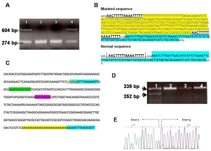
Methods: Routine ophthalmologic examination and direct sequencing of all coding regions of the OPA1 gene were performed. Further characterization of a new OPA1 gene insertion was performed by reverse transcription-PCR (RT-PCR) of RNA from patients and control subjects.
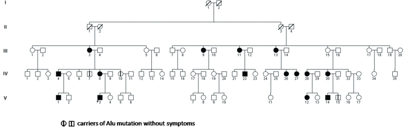
Results: We identified an Alu-element insertion located in intron 7 of OPA1 causing an in-frame deletion of exon 8 in 18 family members.
Conclusions: The predicted consequence of this mutation is the loss of the guanosine triphosphatase (GTPase) activity of OPA1. Alu insertions have been reported in the literature as causing human genetic disease. However, this is the first report of a pathogenic OPA1 gene mutation resulting from an Alu insertion.
Figures
Similar articles
-
First report of OPA1 screening in Greek patients with autosomal dominant optic atrophy and identification of a previously undescribed OPA1 mutation.Mol Vis. 2014 May 27;20:691-703. eCollection 2014. Mol Vis. 2014. PMID: 24883014 Free PMC article.
-
A comprehensive survey of mutations in the OPA1 gene in patients with autosomal dominant optic atrophy.Invest Ophthalmol Vis Sci. 2002 Jun;43(6):1715-24. Invest Ophthalmol Vis Sci. 2002. PMID: 12036970
-
Characterization of two novel intronic OPA1 mutations resulting in aberrant pre-mRNA splicing.BMC Med Genet. 2017 Feb 28;18(1):22. doi: 10.1186/s12881-017-0383-x. BMC Med Genet. 2017. PMID: 28245802 Free PMC article.
-
Meta-analysis of genotype-phenotype analysis of OPA1 mutations in autosomal dominant optic atrophy.Mitochondrion. 2019 May;46:262-269. doi: 10.1016/j.mito.2018.07.006. Epub 2018 Aug 27. Mitochondrion. 2019. PMID: 30165240
-
Recessive optic atrophy, sensorimotor neuropathy and cataract associated with novel compound heterozygous mutations in OPA1.Mol Med Rep. 2016 Jul;14(1):33-40. doi: 10.3892/mmr.2016.5209. Epub 2016 May 4. Mol Med Rep. 2016. PMID: 27150940 Free PMC article. Review.
Cited by
-
Alu insertion variants alter mRNA splicing.Nucleic Acids Res. 2019 Jan 10;47(1):421-431. doi: 10.1093/nar/gky1086. Nucleic Acids Res. 2019. PMID: 30418605 Free PMC article.
-
Retrotransposon-induced mosaicism in the neural genome.Open Biol. 2018 Jul;8(7):180074. doi: 10.1098/rsob.180074. Open Biol. 2018. PMID: 30021882 Free PMC article. Review.
-
Alu mobile elements: from junk DNA to genomic gems.Scientifica (Cairo). 2012;2012:545328. doi: 10.6064/2012/545328. Epub 2012 Dec 16. Scientifica (Cairo). 2012. PMID: 24278713 Free PMC article. Review.
-
Retrotransposon insertion as a novel mutational event in Bardet-Biedl syndrome.Mol Genet Genomic Med. 2019 Feb;7(2):e00521. doi: 10.1002/mgg3.521. Epub 2018 Nov 28. Mol Genet Genomic Med. 2019. PMID: 30484961 Free PMC article.
-
Association of an intronic, but not any exonic, FRMD4B sequence variant and heart failure.Clin Transl Sci. 2010 Aug;3(4):134-9. doi: 10.1111/j.1752-8062.2010.00220.x. Clin Transl Sci. 2010. PMID: 20718813 Free PMC article.
References
-
- Kjer B, Eiberg H, Kjer P, Rosenberg T. Dominant optic atrophy mapped to chromosome 3q region. II. Clinical and epidemiological aspects. Acta Ophthalmol Scand. 1996;74:3–7. - PubMed
-
- Carelli V, Ross-Cisneros FN, Sadun AA. Mitochondrial dysfunction as a cause of optic neuropathies. Prog Retin Eye Res. 2004;23:53–89. - PubMed
-
- Delettre C, Lenaers G, Pelloquin L, Belenguer P, Hamel CP. OPA1 (Kjer type) dominant optic atrophy: a novel mitochondrial disease. Mol Genet Metab. 2002;75:97–107. - PubMed
-
- Frezza C, Cipolat S, Martins de Brito O, Micaroni M, Beznoussenko GV, Rudka T, Bartoli D, Polishuck RS, Danial NN, De Strooper B, Scorrano L. OPA1 controls apoptotic cristae remodeling independently from mitochondrial fusion. Cell. 2006;126:177–89. - PubMed
-
- Puomila A, Huoponen K, Mäntyjärvi M, Hämäläinen P, Paananen R, Sankila EM, Savontaus ML, Somer M, Nikoskelainen E. Dominant optic atrophy: correlation between clinical and molecular genetic studies. Acta Ophthalmol Scand. 2005;83:337–46. - PubMed
Publication types
MeSH terms
Substances
LinkOut - more resources
Full Text Sources
Other Literature Sources