Effects of TGF-beta1 Ribbon Antisense on CCl(4)-induced Liver Fibrosis
- PMID: 20157387
- PMCID: PMC2817527
- DOI: 10.4196/kjpp.2008.12.1.1
Effects of TGF-beta1 Ribbon Antisense on CCl(4)-induced Liver Fibrosis
Abstract
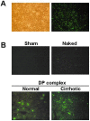
Ribbon-type antisense oligonucleotide to TGF-beta1 (TGF-beta1 RiAS) was designed and tested to prevent or resolve the fibrotic changes induced by CCl4 injection. When Hepa1c1c7 cells were transfected with TGF-beta1 RiAS, the level of TGF-beta1 mRNA was effectively reduced. TGF-beta1 RiAS, mismatched RiAS, and normal saline were each injected to mice via tail veins. When examined for the biochemical effects on the liver, TGF-beta1 mRNA levels were significantly reduced only in the TGF-beta1 RiAS-treated group. The results of immunohistochemical studies showed that TGF-beta1 RiAS prevented the accumulation of collagen and alpha-smooth muscle actin, but could not resolve established fibrosis. These results indicate that ribbon antisense to TGF-beta1 with efficient uptake can effectively prevent fibrosis of the liver.
Keywords: Cationic peptide; Liver cirrhosis; Ribbon antisense; Transforming growth factor-β1.
Figures


Similar articles
-
Prevention of CCl4-induced liver cirrhosis by ribbon antisense to transforming growth factor-beta1.Int J Mol Med. 2008 Jan;21(1):33-9. Int J Mol Med. 2008. PMID: 18097613
-
Prevention of tissue injury by ribbon antisense to TGF-beta1 in the kidney.Int J Mol Med. 2005 Mar;15(3):391-9. Int J Mol Med. 2005. PMID: 15702227
-
Adenoviral expression of a transforming growth factor-beta1 antisense mRNA is effective in preventing liver fibrosis in bile-duct ligated rats.BMC Gastroenterol. 2003 Oct 18;3:29. doi: 10.1186/1471-230X-3-29. BMC Gastroenterol. 2003. PMID: 14565855 Free PMC article.
-
Ghrelin Attenuates Liver Fibrosis through Regulation of TGF-β1 Expression and Autophagy.Int J Mol Sci. 2015 Sep 10;16(9):21911-30. doi: 10.3390/ijms160921911. Int J Mol Sci. 2015. PMID: 26378522 Free PMC article.
-
Adenoviral delivery of an antisense RNA complementary to the 3' coding sequence of transforming growth factor-beta1 inhibits fibrogenic activities of hepatic stellate cells.Cell Growth Differ. 2002 Jun;13(6):265-73. Cell Growth Differ. 2002. PMID: 12114216
Cited by
-
Fucoidan from Fucus vesiculosus protects against alcohol-induced liver damage by modulating inflammatory mediators in mice and HepG2 cells.Mar Drugs. 2015 Feb 16;13(2):1051-67. doi: 10.3390/md13021051. Mar Drugs. 2015. PMID: 25690093 Free PMC article.
-
The Potential Protective Effect of Oligoribonucleotides-d-Mannitol Complexes against Thioacetamide-Induced Hepatotoxicity in Mice.Pharmaceuticals (Basel). 2018 Aug 6;11(3):77. doi: 10.3390/ph11030077. Pharmaceuticals (Basel). 2018. PMID: 30082619 Free PMC article.
References
-
- Alcolado R, Arthur MJ, Iredale JP. Pathogenesis of liver fibrosis. Clin Sci (Lond) 1997;92:103–112. - PubMed
-
- Arias M, Lahme B, Van de Leur E, Gressner AM, Weiskirchen R. Adenoviral delivery of an antisense RNA complementary to the 3' coding sequence of transforming growth factor-beta1 inhibits fibrogenic activities of hepatic stellate cells. Cell Growth Differ. 2002;13:265–273. - PubMed
-
- Arthur MJ, Fibrogenesis II. Metalloproteinases and their inhibitors in liver fibrosis. Am J Physiol Gastrointest Liver Physiol. 2000;279:G245–G249. - PubMed
-
- Bajpai AK, Park JH, Moon IJ, Kang H, Lee YH, Doh KO, Suh SI, Chang BC, Park JG. Rapid blockade of telomerase activity and tumor cell growth by the DPL lipofection of ribbon antisense to hTR. Oncogene. 2005;24:6492–6501. - PubMed

