. 2010 Jan 18:3:1.
doi: 10.1186/1757-2215-3-1.

The prosurvival activity of ascites against TRAIL is associated with a shorter disease-free interval in patients with ovarian cancer

Affiliations

The prosurvival activity of ascites against TRAIL is associated with a shorter disease-free interval in patients with ovarian cancer

Denis Lane et al. J Ovarian Res. .

Abstract

Background: The production of ascites is a common complication of ovarian cancer. Ascites constitute a unique tumor microenvironment that may affect disease progression. In this context, we recently showed that ovarian cancer ascites may protect tumor cells from TRAIL-induced apoptosis. In this study, we sought to determine whether the prosurvival effect of ascites affects disease-free intervals.

Methods: Peritoneal fluids were obtained from 54 women undergoing intra-abdominal surgery for suspected ovarian cancer (44 cancers and 10 benign diseases). The ability of peritoneal fluids to protect from TRAIL was assessed in the ovarian cancer cell line CaOV3, and IC(50 )were determined. The anti-apoptotic activity of 6 ascites against cisplatin, paclitaxel, doxorubicin, etoposide and vinorelbine was also assessed in CaOV3 cells, and the prosurvival activity of two ascites was assessed in 9 primary ovarian cancer cultures.

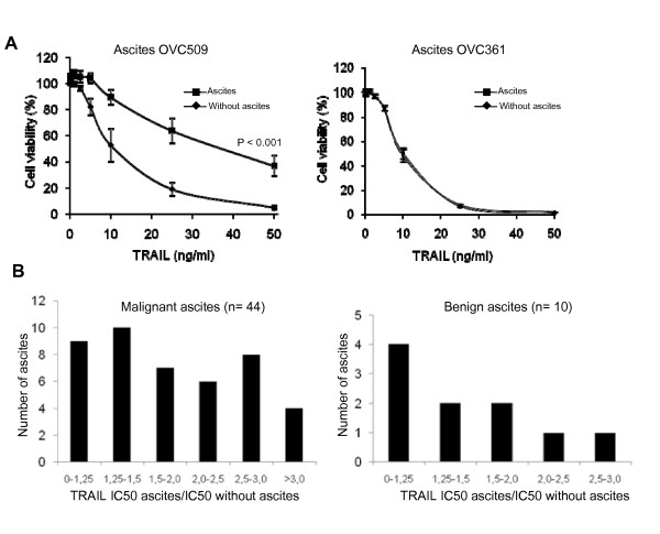
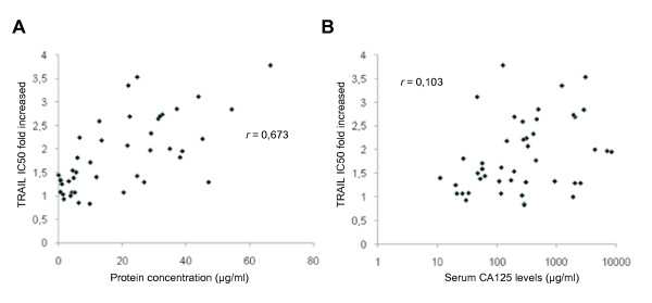
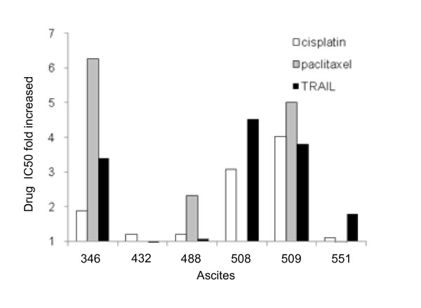
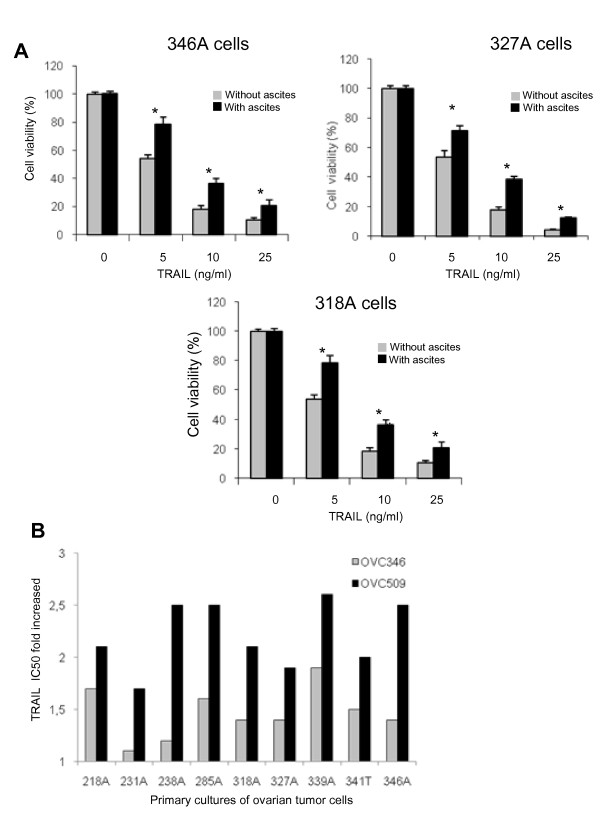
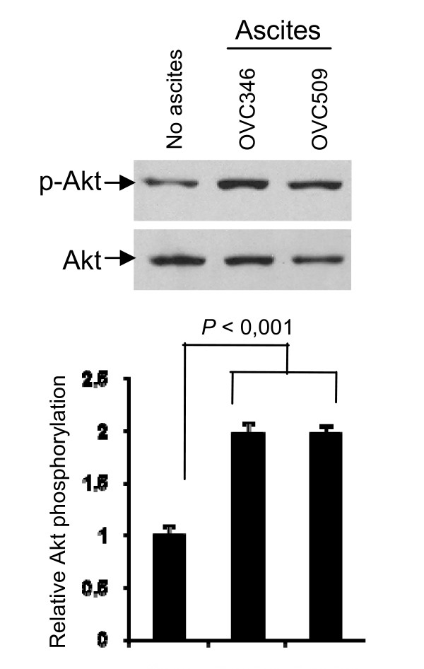
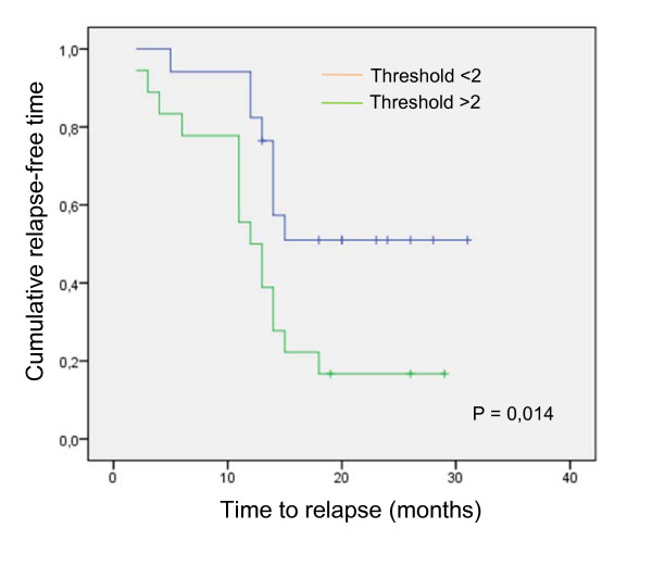
Results: Among the 54 peritoneal fluids tested, inhibition of TRAIL cytotoxicity was variable. Fluids originating from ovarian cancer were generally more protective than fluids from non-malignant diseases. Most of the 44 ovarian cancer ascites increased TRAIL IC(50 )and this inhibitory effect did not correlate strongly with the protein concentration in these ascites or the levels of serum CA125, a tumor antigen which is used in the clinic as a marker of tumor burden. The effect of ascites on cisplatin- and paclitaxel-induced cell death was assessed with 4 ascites having inhibitory effect on TRAIL-induced cell death and 2 that do not. The four ascites with prosurvival activity against TRAIL had some inhibitory on cisplatin and/or paclitaxel. Two ovarian cancer ascites, OVC346 and OVC509, also inhibited TRAIL cytotoxicity in 9 primary cultures of ovarian tumor and induced Akt activation in three of these primary cultures. Among a cohort of 35 patients with ascites, a threshold of TRAIL IC(50 )with ascites/IC(50 )without ascites > 2 was associated with shorter disease-free interval.

Conclusions: The prosurvival activity of ascites against TRAIL is associated with shorter disease-free interval, which may be explained, at least in part, by ascites-induced cisplatin/paclitaxel resistance. Our findings suggest that ascites may contain prosurvival factors that protect against TRAIL and chemotherapy and consequently affect disease progression.


Figures

Figure 1
Figure 1
Effect of peritoneal fluids on TRAIL-induced cell death in CaOV3 cells. (a) CaOV3 cells were pre-incubated for 2 h with OVC509 and OVC361 ascites (10% v/v) obtained from women with advanced serous ovarian cancer and treated with TRAIL (10 ng/ml) for 48 h. Cell viability was measured by XTT assay. Data are shown as the percent cell viability relative to untreated (no TRAIL, no ascites) cells. Results are from three independent experiments done in triplicate and express as mean ± SEM. (b) TRAIL IC50 was determined by XTT assay and defined as the concentration of TRAIL required to kill 50% of CaOV3 cells in the presence or absence of a specific ascites. The prosurvival activity of ovarian cancer ascites and benign fluids was determined by their ability to increase TRAIL IC50 after 48 h compared to the TRAIL IC50 of CaOV3 cells not exposed to peritoneal fluids. A value of 1 indicates a neutral effect of ascites on TRAIL-induced cytoxicity.
Figure 2
Figure 2
Protein concentration of peritoneal fluids and baseline serum CA125 levels. (a) Protein concentration of the 44 ovarian cancer ascites was determined and correlated with TRAIL IC50 fold increased mediated by ascites. (b) Baseline serum CA125 levels were obtained for all except one patient and correlated with TRAIL IC50 fold increased mediated by ascites. Correlation coefficients (r) were determined by Pearson's correlation coefficient test.
Figure 3
Figure 3
Effect of ovarian cancer ascites on TRAIL-, cisplatin- and paclitaxel-induced cell death in CaOV3 cells. CaOV3 cells were pre-incubated for 2 h with various fluids (10% v/v) obtained from women with advanced ovarian cancer and treated with increasing concentrations of TRAIL for 48 h or with cisplatin or paclitaxel for 72 h. Cell viability was assessed by XTT assays. TRAIL, cisplatin and paclitaxel IC50 were determined in the presence of ascites and expressed as fold increased relative to IC50 in the absence of ascites. A value of 1 indicates a neutral effect of ascites on these drugs. Results are from three independent experiments done in triplicate.
Figure 4
Figure 4
Effect of ovarian cancer ascites on TRAIL-induced cell death in primary ovarian tumor samples. (A) Primary cultures ovarian tumor cells (samples 346, 327, 318) were pre-incubated for 2 h with OVC509 (10% v/v) and treated with increasing TRAIL concentrations for 48 h. Cell viability was measured by XTT assay. Data are shown as the percent cell viability relative to TRAIL and ascites untreated cells. Results are from three independent experiments done in triplicate and express as mean ± SEM. *, indicates P < 0,001. (b) TRAIL IC50 were determined in the presence of OVC346 or OVC509 ascites and expressed as fold increased relative to IC50 in the absence of ascites for 9 primary cultures of ovarian tumor cells. Cells were isolated either from ascites (A) or from tissues (T). A value of 1 indicates a neutral effect of ascites on TRAIL cytotoxicity.
Figure 5
Figure 5
Ovarian cancer ascites OVC346 and OVC509 were incubated with primary cultures from sample 346A for 90 min. Lysates were obtained and Western blot analysis was performed with phospho-Ser473 Akt (p-Akt) and Akt antibody (Akt). Densitometric quantification of phosphorylated Akt from three separate experiments normalized to total Akt. Data are expressed as Akt phosphorylation fold increased relative to 349A cells not treated with ascites.
Figure 6
Figure 6
Impact of having protective ascites on time to first relapse. Kaplan-Meier curve for 35 patients with ovarian cancer ascites showing the association between protective or non-protective ascites and disease-free interval. Log-rank test was used to verify the significance of the difference (P = 0.014).

References

    1. Jemal A, Siegel R, Ward E, Hao Y, Xu J, Murray T, Thun MJ. Cancer statistics, 2008. CA Cancer J Clin. 2008;49:8–31. - PubMed
    1. Auersperg N, Ota T, Mitchell GW. Early events in ovarian epithelial carcinogenesis: progress and problems in experimental approaches. Int J Gynecol Cancer. 2002;12:691–703. doi: 10.1046/j.1525-1438.2002.01152.x. - DOI - PubMed
    1. Mills GB, May C, McGill M, Roifman CM, Mellors A. A putative new growth factor in ascitic fluid from ovarian cancer patients: identification, characterization, and mechanism of action. Cancer Res. 1988;48:1066–1071. - PubMed
    1. Mills GB, May C, Hill M, Campbell S, Shaw P, Marks A. Ascitic fluid from human ovarian cancer patients contains growth factors necessary for intraperitoneal growth of human ovarian adenocarcinoma cells. J Clin Invest. 1990;86:851–855. doi: 10.1172/JCI114784. - DOI - PMC - PubMed
    1. Xu Y, Gaudette DC, Boynton JD, Frankel A, Fang XJ, Sharma A, Hurteau J, Casey G, Goodbody A, Mellors A, Holub BJ, Mills GB. Characterization of an ovarian cancer activating factor in ascites of ovarian cancer patients. Clin Cancer Res. 1995;1:1223–1232. - PubMed