. 2010 Jan 6:7:1.
doi: 10.1186/1743-8454-7-1.

The regulation of brain states by neuroactive substances distributed via the cerebrospinal fluid; a review

Affiliations

The regulation of brain states by neuroactive substances distributed via the cerebrospinal fluid; a review

Jan G Veening et al. Cerebrospinal Fluid Res. .

Abstract

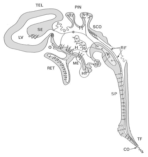
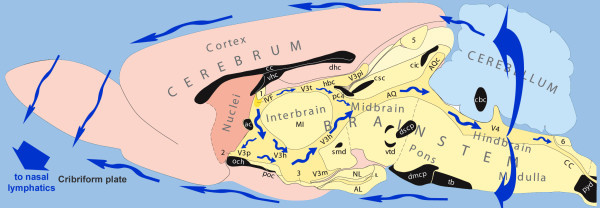
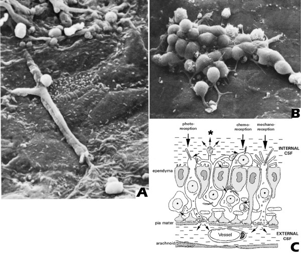
The cerebrospinal fluid (CSF) system provides nutrients to and removes waste products from the brain. Recent findings suggest, however, that in addition, the CSF contains message molecules in the form of actively released neuroactive substances. The concentrations of these vary between locations, suggesting they are important for the changes in brain activity that underlie different brain states, and induce different sensory input and behavioral output relationships.The cranial CSF displays a rapid caudally-directed ventricular flow followed by a slower rostrally-directed subarachnoid flow (mainly towards the cribriform plate and from there into the nasal lymphatics). Thus, many brain areas are exposed to and can be influenced by substances contained in the CSF. In this review we discuss the production and flow of the CSF, including the mechanisms involved in the regulation of its composition. In addition, the available evidence for the release of neuropeptides and other neuroactive substances into the CSF is reviewed, with particular attention to the selective effects of these on distant downstream receptive brain areas. As a conclusion we suggest that (1) the flowing CSF is involved in more than just nutrient and waste control, but is also used as a broadcasting system consisting of coordinated messages to a variety of nearby and distant brain areas; (2) this special form of volume transmission underlies changes in behavioral states.


Figures

Figure 1
Figure 1
A schematic diagram of structures and specialized cell types bordering the different parts of the mammalian ventricular system, and in contact with the cerebrospinal fluid (CSF). The complexity of the system suggests that CSF functions are not limited to metabolic support of the brain and the release of waste products. Abbreviations: CO: caudal opening of the central canal of the spinal cord;H: hypothalamic CSF-contacting neurons; HY: Hypophysis; LV: lateral ventricle; ME: median eminence; O: vascular organ of the terminal lamina; PIN: pineal organ; R: raphe nuclei; RET: retina; RF: Reissner's fiber; SE: septal region; SCO: subcommissural organ; SP: medullo-spinal CSF-contacting neurons; TEL: telencephalon; TF: terminal filum; (Fig. 1 was kindly provided by Prof. B. Vigh. For details about specific cell types, the reader is referred to: Vigh and Vigh-Teichmann, [31], and to Vigh et al, [32]).
Figure 2
Figure 2
A schematic diagram showing the direction of the CSF bulk flow in the mammalian brain based on a midsagittal section of the rat brain, kindly provided by Prof. L.W. Swanson. The following regions are indicated: telencephalon (pink shades); diencephalon (interbrain) and brainstem areas (yellow shades); cerebellum dorsal to the brainstem (light blue); sectioned fiber tracts (black). CSF (blue arrows) flows from the lateral ventricles to the third ventricle via the interventricular foramen (IVF), and flows caudally along the dorsal and ventral side of thalamic adhesion, to the cerebral aqueduct (AQ) and fourth ventricle (V4). Some CSF may continue flowing caudally through the central canal of the spinal cord (CC), but most leaves the ventricular system via the lateral apertures and flows through the subarachnoid space, surrounding the brain. This external flow is indicated here along the dorsal and ventral side of the brain but occurs also along all other external brain surfaces. The destination of the subarachnoid flow is the cribriform plate of the ethmoidal bone, containing the penetrating olfactory fibers, where CSF is released in small lymphatic vessels. Additional abbreviations: V3(p, h, m, t, pi): regions and recesses of the third ventricle; Circumventricular organs: 1: subfornical organ; 2: organum vasculosum of the lamina terminalis; 3: median eminence; 4: subcommissural organ; 5: pineal organ; 6: area postrema; AL, IL and NL: different lobes of pituitary; Fiber bundles crossing the midline, coloured black, are not relevant for the present review.
Figure 3
Figure 3
(A and B) show scanning electron microscopy of supraependymal structures in the rat brain. A: Partially ensheathed fibers and terminal arborizations; B: cellular structures with short fiber extensions, on the ventricular surface of the ependymal cells; Fig 3C: a schematic diagram of the ependymal layer with supra- and subependymal cellular structures contacting the CSF at the ventricular side of the layer; the * indicates a supraependymal terminal extending into the CSF, as described for serotonergic fibers. Figs 3A and 3B were reprinted from [121], with permission of Springer-Verlag. Fig 3C was kindly provided by Prof. B. Vigh.

Similar articles

Cited by

References

    1. Frijda N. The laws of emotions. London: Psychology Press; 2007.
    1. Hinde RA. Animal Behaviour; A Synthesis of Ethology and Comparative Psychology. second. New York: McGRAW-Hill Book Cy; 1970.
    1. Mogenson GJ. The neurobiology of Behavior: an introduction. Hillsdale: Lawrence Erlbaum Associates; 1977.
    1. Panksepp J. Affective Neuroscience, The foundations of human and animal emotions. New York: Oxford University Press; 1998.
    1. Swanson LW. Brain Architecture, understanding the basic plan. New York: Oxford University Press; 2003.