

Correlations between capnographic waveforms and peak flow meter measurement in emergency department management of asthma
- PMID: 20157449
- PMCID: PMC2700227
- DOI: 10.1007/s12245-009-0088-9
Correlations between capnographic waveforms and peak flow meter measurement in emergency department management of asthma
Abstract
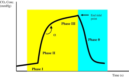
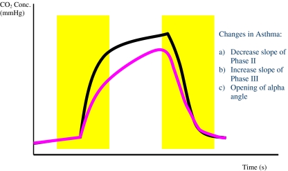
Background: The usual method for initial assessment of an acute asthma attack in the emergency room includes the use of peak flow measurement and clinical parameters. Both methods have their own disadvantages such as poor cooperation/effort from patients (peak flow meter) and lack of objective assessment (clinical parameters). We were looking into other methods for the initial asthma assessment, namely the use of capnography. The normal capnogram has an almost square wave pattern comprising phase 1, slope phase 2, plateau phase 3, phase 4 and angle alpha (between slopes 2 and 3). The changes in asthma include decrease in slope of phase 2, increase in slope 3 and opening of angle alpha.
Aims: Our objective was to compare and assess the correlation between the changes in capnographic indices and peak flow measurement in non-intubated acute asthmatic patients attending the emergency room.
Methods: We carried out a prospective study in a university hospital emergency department (ED). One hundred and twenty eight patients with acute asthma were monitored with peak flow measurements and then had a nasal cannula attached for microstream sampling of expired carbon dioxide. The capnographic waveform was recorded onto a PC card for indices analysis. The patients were treated according to departmental protocols. After treatment, when they were adjudged well for discharge, a second set of results was obtained for peak flow measurements and capnographic waveform recording. The pre-treatment and post-treatment results were then compared with paired samples t-test analysis. Simple and canonical correlations were performed to determine correlations between the assessment methods. A p value of below 0.05 was taken to be significant.
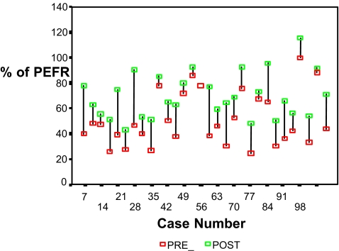
Results: Peak flow measurements showed significant improvements post-treatment (p < 0.001). On the capnographic waveform, there was a significant difference in the slope of phase 3 (p < 0.001) and alpha angle (p < 0.001), but not in phase 2 slope (p = 0.35). Correlation studies done between the assessment methods and indices readings did not show strong correlations either between the measurements or the magnitude of change pre-treatment and post-treatment.
Conclusion: Peak flow measurements and capnographic waveform indices can indicate improvements in airway diameter in acute asthmatics in the ED. Even though the two assessment methods did not correlate statistically, capnographic waveform analysis presents several advantages in that it is effort independent and provides continuous monitoring of normal tidal respiration. They can be proposed for the monitoring of asthmatics in the ED.
Keywords: Asthma; Capnography; Emergency room; Peak flow measurement.
Figures
References
-
- O’Flaherty D. Capnography: principles and practice. London: BMJ Publishing Group; 1994.
-
- Smalhout B, Kalenda Z. An atlas of capnography. Amsterdam: Kerchebosch-Zeist; 1981.
-
- Berrengo A, Cutilloa A. Single-breath analysis of carbon dioxide concentration records. J Appl Physiol. 1961;16:522–530.
-
- You B, Mayeux D, Rkiek B, Autran N, Dang Vu V, Grilliat JP. La capnographie expiratoire dans l’asthme: perspectives d’utilisation comme methode de monitorage. Rev Mal Respir. 1992;9:547–552. - PubMed
LinkOut - more resources
Full Text Sources
