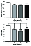
Is thymocyte development functional in the aged?
- PMID: 20157506
- PMCID: PMC2806005
- DOI: 10.18632/aging.100027
Is thymocyte development functional in the aged?
Abstract
T cells are an integral part of a functional immune system with the majority being produced in the thymus. Of all the changes related to immunosenescence, regression of the thymus is considered one of the most universally recognised alterations. Despite the reduction of thymic size, there is evidence to suggest that T cell output is still present into old age, albeit much diminished; leading to the assumption that thymocyte development is normal. However, current data suggests that recent thymic emigrant from the aged thymus are functionally less responsive, giving rise to the possibility that the generation of naïve T cell may be intrinsically impaired in the elderly. In light of these findings we discuss the evidence that suggest aged T cells may be flawed even before exiting to the periphery and could contribute to the age-associated decline in immune function.
Keywords: T cells; aging; immunity; thymocyte; thymus.
Conflict of interest statement
The authors in this manuscript have no conflict of interests to declare.
Figures


Similar articles
-
Flow cytometric analysis of CD3/TCR complex, zinc, and glucocorticoid-mediated regulation of apoptosis and cell cycle distribution in thymocytes from old mice.Cytometry. 1998 May 1;32(1):1-8. Cytometry. 1998. PMID: 9581618
-
Luteinizing hormone-releasing hormone (LHRH) agonist restoration of age-associated decline of thymus weight, thymic LHRH receptors, and thymocyte proliferative capacity.Endocrinology. 1989 Aug;125(2):1037-45. doi: 10.1210/endo-125-2-1037. Endocrinology. 1989. PMID: 2546733
-
Development and functional properties of thymic and extrathymic T lymphocytes.Crit Rev Immunol. 2008;28(5):441-66. doi: 10.1615/critrevimmunol.v28.i5.40. Crit Rev Immunol. 2008. PMID: 19166388 Review.
-
Thymic regeneration: teaching an old immune system new tricks.Trends Mol Med. 2002 Oct;8(10):469-76. doi: 10.1016/s1471-4914(02)02415-2. Trends Mol Med. 2002. PMID: 12383769 Review.
-
Involution of the mammalian thymus, one of the leading regulators of aging.In Vivo. 1997 Sep-Oct;11(5):421-40. In Vivo. 1997. PMID: 9427047
Cited by
-
Changes in the frequencies of human hematopoietic stem and progenitor cells with age and site.Exp Hematol. 2014 Feb;42(2):146-54. doi: 10.1016/j.exphem.2013.11.003. Epub 2013 Nov 15. Exp Hematol. 2014. PMID: 24246745 Free PMC article.
-
The origin and implication of thymic involution.Aging Dis. 2011 Oct;2(5):437-43. Epub 2011 Oct 28. Aging Dis. 2011. PMID: 22396892 Free PMC article.
-
Senescence-accelerated OXYS rats: a model of age-related cognitive decline with relevance to abnormalities in Alzheimer disease.Cell Cycle. 2014;13(6):898-909. doi: 10.4161/cc.28255. Epub 2014 Feb 17. Cell Cycle. 2014. PMID: 24552807 Free PMC article. Review.
-
Anidulafungin Versus Micafungin in the Treatment of Candidemia in Adult Patients.Mycopathologia. 2020 Aug;185(4):653-664. doi: 10.1007/s11046-020-00471-8. Epub 2020 Jul 23. Mycopathologia. 2020. PMID: 32705415 Free PMC article.
-
Mitochondria-targeted antioxidant SkQ1 inhibits age-dependent involution of the thymus in normal and senescence-prone rats.Aging (Albany NY). 2009 Apr 22;1(4):389-401. doi: 10.18632/aging.100043. Aging (Albany NY). 2009. PMID: 20195490 Free PMC article.
References
-
- Pawelec G, Akbar A, Caruso C, Solana R, Grubeck-Loebenstein B, Wikby A. Human immunosenescence: is it infectious. Immunol Rev. 2005;205:257–268. - PubMed
-
- Aspinall R, Andrew D. Thymic atrophy in the mouse is a soluble problem of the thymic environment. Vaccine. 2000;18:1629–1637. - PubMed
-
- Aw D, Silva AB, Maddick M, von Zglinicki T, Palmer DB. Architectural changes in the thymus of aging mice. Aging Cell. 2008;7:158–167. - PubMed
-
- George AJ, Ritter MA. Thymic involution with ageing: obsolescence or good housekeeping. Immunol Today. 1996;17:267–272. - PubMed
Publication types
MeSH terms
Substances
Grants and funding
LinkOut - more resources
Full Text Sources
Medical
