

Serum markers of apoptosis decrease with age and cancer stage
- PMID: 20157546
- PMCID: PMC2806040
- DOI: 10.18632/aging.100069
Serum markers of apoptosis decrease with age and cancer stage
Abstract
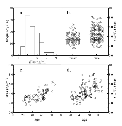
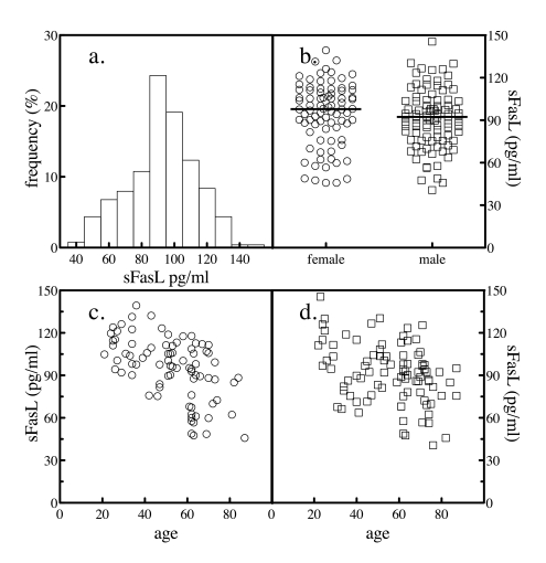
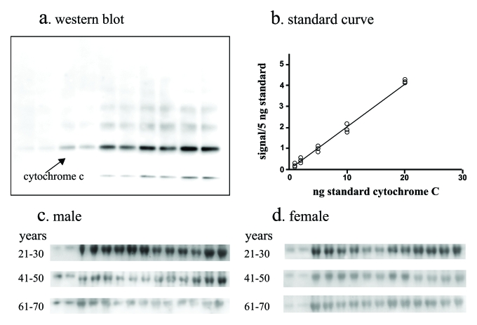
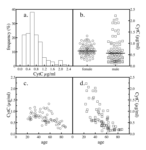
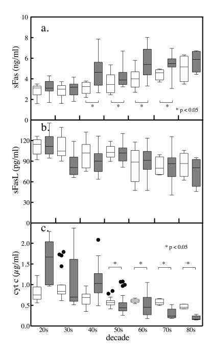
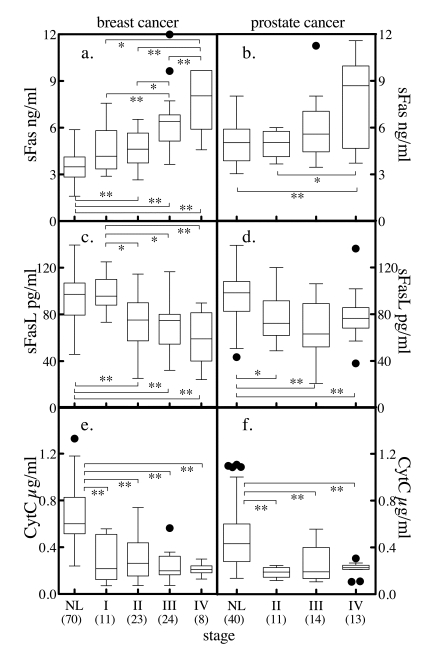
The physical manifestations of aging reflect a loss of homeostasis that effects molecular, cellular and organ system functional capacity. As a sentinel homeostatic pathway, changes in apoptosis can have pathophysiological consequences in both aging and disease. To assess baseline global apoptosis balance, sera from 204 clinically normal subjects had levels of sFas (inhibitor of apoptosis), sFasL (stimulator of apoptosis), and total cytochrome c (released from cells during apoptosis) measured. Serum levels of sFas were significantly higher while sFasL and cytochrome c levels were lower in men compared to women. With increasing age there was a decrease in apoptotic markers (cytochrome c) and pro-apoptotic factors (sFasL) and an increase in anti-apoptotic factors (sFas) in circulation. The observed gender differences are consistent with the known differences between genders in mortality and morbidity. In a separate cohort, subjects with either breast (n = 66) or prostate cancer (n = 38) exhibited significantly elevated sFas with reduced sFasL and total cytochrome c regardless of age. These markers correlated with disease severity consistent with tumor subversion of apoptosis. The shift toward less global apoptosis with increasing age in normal subjects is consistent with increased incidence of diseases whose pathophysiology involves apoptosis dysregulation.
Keywords: aging; apoptosis; cancer; cytochrome c; immunosenescence; serum markers.
Conflict of interest statement
There is no conflict of interest for any of the authors.
Figures
Similar articles
-
Matrix metalloproteinases and soluble Fas/FasL system as novel regulators of apoptosis in children and young adults on chronic dialysis.Apoptosis. 2011 Jul;16(7):653-9. doi: 10.1007/s10495-011-0604-2. Apoptosis. 2011. PMID: 21516345 Free PMC article.
-
Effect of tibolone and raloxifene on serum markers of apoptosis in postmenopausal women.Climacteric. 2013 Apr;16(2):258-64. doi: 10.3109/13697137.2012.668251. Epub 2012 May 29. Climacteric. 2013. PMID: 22642937 Clinical Trial.
-
Soluble Fas/FasLare elevated in the serum and cerebrospinal fluid of patients with neurocysticercosis.Parasitol Res. 2017 Nov;116(11):3027-3036. doi: 10.1007/s00436-017-5613-9. Epub 2017 Sep 30. Parasitol Res. 2017. PMID: 28965226
-
Apoptosis Markers in Breast Cancer Therapy.Adv Clin Chem. 2016;74:143-93. doi: 10.1016/bs.acc.2015.12.003. Epub 2016 Jan 19. Adv Clin Chem. 2016. PMID: 27117663 Review.
-
CSF soluble Fas correlates with the severity of HIV-associated dementia.Neurology. 2004 Feb 24;62(4):654-6. doi: 10.1212/01.wnl.0000110188.37546.51. Neurology. 2004. PMID: 14981191 Review.
Cited by
-
Low FasL levels promote proliferation of human bone marrow-derived mesenchymal stem cells, higher levels inhibit their differentiation into adipocytes.Cell Death Dis. 2013 Apr 18;4(4):e594. doi: 10.1038/cddis.2013.115. Cell Death Dis. 2013. PMID: 23598406 Free PMC article.
-
Role of apoptosis in disease.Aging (Albany NY). 2012 May;4(5):330-49. doi: 10.18632/aging.100459. Aging (Albany NY). 2012. PMID: 22683550 Free PMC article. Review.
-
Distinct roles of trauma and transfusion in induction of immune modulation after injury.Transfusion. 2012 Dec;52(12):2533-50. doi: 10.1111/j.1537-2995.2012.03618.x. Epub 2012 Mar 27. Transfusion. 2012. PMID: 22452342 Free PMC article.
-
Sex-related differences in urinary immune-related metabolic profiling of alopecia areata patients.Metabolomics. 2020 Jan 16;16(2):15. doi: 10.1007/s11306-020-1634-y. Metabolomics. 2020. PMID: 31950279
-
Programmed cell death in aging.Ageing Res Rev. 2015 Sep;23(Pt A):90-100. doi: 10.1016/j.arr.2015.04.002. Epub 2015 Apr 8. Ageing Res Rev. 2015. PMID: 25862945 Free PMC article. Review.
References
-
- Muradian K, Schachtschabel DO. The role of apoptosis in aging and age-related disease: update. Z Gerontol Geriatr. 2001;34:441–446. - PubMed
-
- Pollack M, Phaneuf S, Dirks A, Leeuwenburgh C. The role of apoptosis in the normal aging brain, skeletal muscle, and heart. Ann N Y Acad Sci. 2002;959:93–107. - PubMed
-
- Adams JD, Mukherjee SK, Klaidman LK, Chang ML, Yasharel R. Apoptosis and oxidative stress in the aging brain. Ann N Y Acad Sci. 1996;786:135–151. - PubMed
-
- Sastre J, Pallardo FV, Vina J. Mitochondrial oxidative stress plays a key role in aging and apoptosis. IUBMB Life. 2000;49:427–435. - PubMed
-
- Kujoth GC, Hiona A, Pugh TD, Someya S, Panzer K, Wohlgemuth SE, Hofer T, Seo AY, Sullivan R, Jobling WA, Morrow JD, Van Remmen H. Mitochondrial DNA mutations, oxidative stress, and apoptosis in mammalian aging. Science. 2005;309:481–484. - PubMed
Publication types
MeSH terms
Substances
Grants and funding
LinkOut - more resources
Full Text Sources
Other Literature Sources
Medical
Research Materials