. 2010 Mar;75(6):1539-49.
doi: 10.1111/j.1365-2958.2010.07070.x. Epub 2010 Feb 10.

The IbpA and IbpB small heat-shock proteins are substrates of the AAA+ Lon protease

Affiliations

The IbpA and IbpB small heat-shock proteins are substrates of the AAA+ Lon protease

Sarah A Bissonnette et al. Mol Microbiol. 2010 Mar.

Abstract

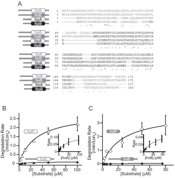
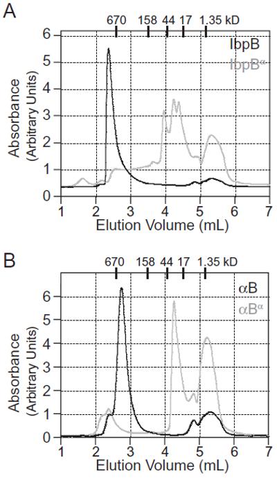
Small heat-shock proteins (sHSPs) are a widely conserved family of molecular chaperones, all containing a conserved alpha-crystallin domain flanked by variable N- and C-terminal tails. We report that IbpA and IbpB, the sHSPs of Escherichia coli, are substrates for the AAA+ Lon protease. This ATP-fueled enzyme degraded purified IbpA substantially more slowly than purified IbpB, and we demonstrate that this disparity is a consequence of differences in maximal Lon degradation rates and not in substrate affinity. Interestingly, however, IbpB stimulated Lon degradation of IbpA both in vitro and in vivo. Furthermore, although the variable N- and C-terminal tails of the Ibps were dispensable for proteolytic recognition, these tails contain critical determinants that control the maximal rate of Lon degradation. Finally, we show that E. coli Lon degrades variants of human alpha-crystallin, indicating that Lon recognizes conserved determinants in the folded alpha-crystallin domain itself. These results suggest a novel mode for Lon substrate recognition and provide a highly suggestive link between the degradation and sHSP branches of the protein quality-control network.


Figures

Figure 1
Figure 1
E. coli sHSPs IbpA and IbpB are Lon substrates A. Sequence alignment of IbpA and IbpB generated by ClustalW2. Darker letters represent the conserved α-crystallin domain. Identical residues are denoted by (*), residues with the same size and hydropathy are denoted by (:), residues with the same size or hydropathy are denoted by (.). B. Lon (600 nM hexamer) degradation of 5 μM IbpA (0.010 ± 0.001 min−1 Lon6−1) or 5 μM IbpB (0.20 ± 0.02 min−1 Lon6−1). C. Substrate dependence of Lon degradation of IbpA (Vmax = 0.043 ± 0.018 min−1 Lon6−1, KM = 18 ± 16 μM) or IbpB (Vmax = 0.60 ± 0.06 min−1 Lon6−1, KM = 16 ± 2.7 μM). Degradation rates were measured from experiments like the one shown in (B) and were fit to the Michaelis-Menten equation. Error bars (±1 SD) in this and all other figures were calculated from at least three independent experiments. The large error in the KM for IbpA is due to the slow rate of degradation. Inset: The IbpA data are replotted on an expanded scale to show the curvature of the fitted line.
Figure 2
Figure 2
Lon degrades the isolated α-crystallin domains of IbpA and IbpB. A. Degradation of 5 μM substrate by 600 nM Lon6. IbpA (filled squares), IbpB (filled circles), IbpAα (open squares) or IbpBα (open circles). B. Michaelis-Menten plot for IbpAα (Vmax = 0.47 ± 0.06 min−1 Lon6−1, KM = 17 ± 4.7 μM). C. Michaelis-Menten plot for IbpBα (Vmax = 0.19 ± 0.01 min−1 Lon6−1, KM = 16 ± 2.4 μM). For comparison, the plots for IbpA and IbpB are shown in grey.
Figure 3
Figure 3
Lon degrades human α-crystallin proteins. A. Sequence alignment of E. coli IbpA and IbpB and human αA-crystallin and αB-crystallin generated by ClustalW2. B. Michaelis-Menten plots for Lon degradation of full-length αA-crystallin (Vmax = 0.12 ± 0.05 min−1 Lon6−1, KM = 50 ± 20 μM) or αAα (Vmax = 2.5 ± 0.3 min−1 Lon6−1, KM = 35 ± 8.9 μM). C. Michaelis-Menten plots for Lon degradation of full-length αB-crystallin (Vmax = 0.06 ± 0.02 min−1 Lon6−1, KM = 58 ± 21 μM) or αBα (Vmax = 3.8 ± 1.4 min−1 Lon6−1, KM = 26 ± 12 μM). Insets: The data for hαA and hαB are replotted on expanded scales.
Figure 4
Figure 4
Tail-less E. coli IbpB and human αB-crystallin elute as smaller oligomers than their full-length counterparts. Gel-filtration chromatography of purified proteins monitored by absorbance at 280 nm or 213 nm. A. E. coli IbpB (black line) and IbpBα (grey line). B. Human αB-crystallin (black line) and αBα (grey line). The calculated monomer molecular weights of IbpB, IbpBα, αB and αBα are ~16 kDa, ~10 kDa, ~20 kDa and ~10 kDa respectively. Tick marks at the top of each panel indicate the elution positions of molecular-weight standards.
Figure 5
Figure 5
IbpB facilitates IbpA degradation both in vitro and in vivo. A. Degradation of 5 μM 35S-IbpA (grey squares), 5 μM 35S-IbpB (grey circles), 5 μM 35S-IbpA with 5 μM unlabelled IbpB (black squares), and 5 μM 35S-labelled IbpB with 5 μM unlabelled IbpA (black circles) by 600 nM Lon6. Asterisks denote 35S-labelled protein. B. Western blots probed with an IbpA-specific antibody showing the time-course of IbpA degradation in wild-type (top panel, left side), lon (top panel, right side), ibpB (bottom panel, left side) or ibpBlon (bottom panel, right side) strains after inhibiting translation with spectinomycin. Representative blots from one of three independent experiments are shown. C. Bands from the experiments in panel B were quantified, and relative intensities are plotted.
Figure 6
Figure 6
Models for Lon recognition and possible roles for Ibp degradation. A. Lon recognizes the α-crystallin domains of IbpA and IbpB and not the N- and C-terminal tails of these proteins. B. Potential roles for Lon degradation of Ibp proteins in vivo. Lon may degrade free Ibps (pathway 1), degrade client proteins and bound Ibps at the same time (pathway 2) or degrade client-bound Ibps to allow refolding of client proteins (pathway 3).

Similar articles

Cited by

References

    1. Ahner A, Nakatsukasa K, Zhang H, Frizzell RA, Brodsky JL. Small heat-shock proteins select deltaF508-CFTR for endoplasmic reticulum-associated degradation. Mol Biol Cell. 2007;18:806–814. - PMC - PubMed
    1. Allen SP, Polazzi JO, Gierse JK, Easton AM. Two novel heat shock genes encoding proteins produced in response to heterologous protein expression in Escherichia coli. J Bacteriol. 1992;174:6938–6947. - PMC - PubMed
    1. Baker TA, Sauer RT. ATP-dependent proteases of bacteria: recognition logic and operating principles. Trends Biochem Sci. 2006;31:647–653. - PMC - PubMed
    1. Choy JS, Aung LL, Karzai AW. Lon protease degrades transfer-messenger RNA-tagged proteins. J Bacteriol. 2007;189:6564–6571. - PMC - PubMed
    1. Doyle SM, Hoskins JR, Wickner S. Collaboration between the ClpB AAA+ remodeling protein and the DnaK chaperone system. Proc Natl Acad Sci USA. 2007;104:11138–11144. - PMC - PubMed

Publication types

MeSH terms

LinkOut - more resources