

doi: 10.1186/1471-2164-11-S1-S14.
Bioinformatic search of plant microtubule-and cell cycle related serine-threonine protein kinases
Affiliations
- PMID: 20158871
- PMCID: PMC2822528
- DOI: 10.1186/1471-2164-11-S1-S14
Item in Clipboard
Bioinformatic search of plant microtubule-and cell cycle related serine-threonine protein kinases
BMC Genomics.
.
Abstract
A bioinformatic search was carried for plant homologues of human serine-threonine protein kinases involved in regulation of cell division and microtubule protein phosphorylation (SLK, PAK6, PAK7, MARK1, MAST2, TTBK1, TTBK2, AURKA, PLK1, PLK4 and PASK). A number of SLK, MAST2 and AURKA plant homologues were identified. The closest identified homologue of human AURKA kinase was a protein of unknown function, A7PY12/GSVIVT00026259001 from Vitis vinifera (herein named as "STALK", Serine-Threonine Aurora-Like Kinase). Analysis of STALK's three-dimensional structure confirmed its relationship to the subgroup of AURKA-like protein kinases.
Figures
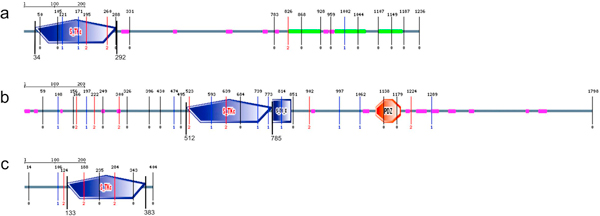
SLK_HUMAN (a), MAST_HUMAN (b) and STK6_HUMAN (c) kinase domain architecture. S_TKc - catalytic domain of serine/threonine-kinases; S_TK_X - auxiliary S_TKc domain; DUF1908 - domain of unknown function (DUF1908); PDZ (also referred as DHR (Dlg homology region) or GLGF (relatively well conserved tetrapeptide in these domains) - domain found in PSD-95, Dlg and ZO-1/2[74]. These domains help anchor transmembrane proteins to the cytoskeleton and hold together signaling complexes [72].
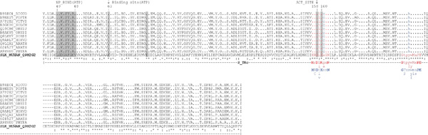
Catalytic domain alignment of human SLK_HUMAN (Q9H2G2) and putative plant SLK-like proteins. NP_BIND (ATP) - nucleotide phosphate binding region; Binding site (ATP) - ATP binding site; ACT_SITE - proton acceptor; consensus conserved motif in catalytic loop region of the subdomain VIb (animals (red): H-R-D-[LI]-K-[GA]-x-N and A. thaliana (blue): H-[RC]-D-[ILV]-K-x-x-N); consensus conserved motif in activation loop of the subdomain VIII (in animals (red): G-T-P-[YF]-[WY]-M-A-P-E and in A. thaliana (blue): G-[TS]-x-x-[WYF]-[ML]-[AS]-P-E)
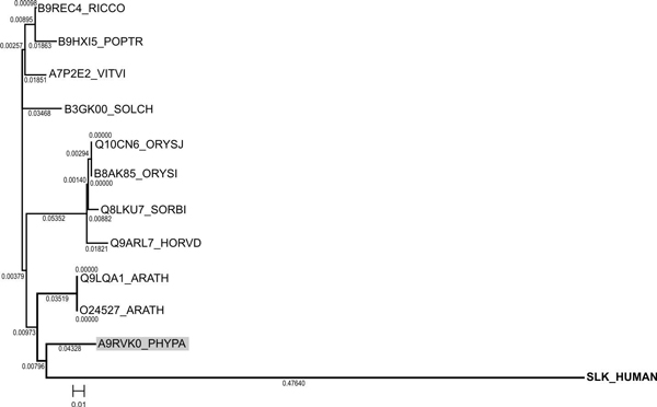
Phylogenetic tree constructed for SLK kinase of H. sapiens and its plant homologues.
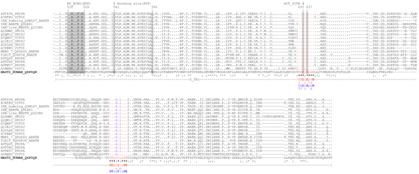
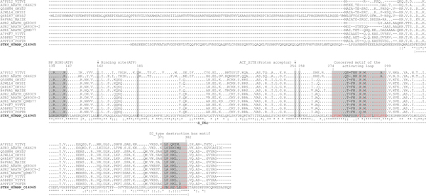
Catalytic domain alignment of MAST_HUMAN (Q6P0Q8) with its plant homologues. NP_BIND (ATP) - nucleotide phosphate binding region; Binding site (ATP) - ATP binding site; ACT_SITE - proton acceptor; consensus conserved for AGC kinases motif in catalytic loop region of the subdomain VIb (animals (red): [HY]-R-D-[LI]-K-[PL]-[ED]-N and A. thaliana (blue): [HY]-[RY]-D-[LI]-K-P-[ED]-N); consensus conserved for AGC kinases motif in activation loop of the subdomain VIII (in animals (red): G-T-P-[EA]-Y-[IM]-A-P-E and in A. thaliana (blue): G-T-x-D-Y-L-A-P-E)
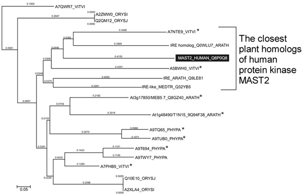
Phylogenetic tree built for H. sapiens MAST2 kinase and its plant homologues; * - plant homologues containing typical for animal MAST2 kinases auxiliary S_TK_X catalytic S_TK_X domain.
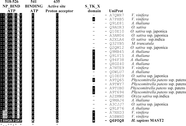
Similarity of H. sapiens MAST2 and potential plant homologues for auxiliary S_TK_X domain, conserved active site Asp-635 and ATP-binding motifs (NP_BIND ATP and Lys-541).
Alignment of human Aurora A and its plant homologues. NP_BIND (ATP) - nucleotide phosphate binding site; Binding site (ATP) - ATP binding site; ACT_SITE - proton acceptor site
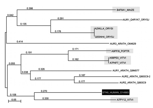
Phylogenetic tree of STK6_HUMAN (Aurora A) kinase and its plant homologues demonstrating their phylogenetic relationships. Highlighted are the proteins identified in the paper.
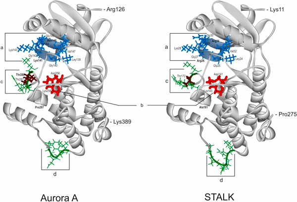
Comparison of the catalytic domain spatial structures of the human protein kinase Aurora A (AURKA, STK6, PDB: 2J4Z) and the protein of unknown function STALK (S_T AURKA LIKE KINASE, UniProt: A7PY12) from V. vinifera. "a" (marked by blue) ATP-binding regions in Aurora A and STALK; "b" (marked by red) is active site; "c", "d" (marked by green) are the most spatially variable regions between the two proteins; phosphorylated Thr residues (287, 288) in the Aurora A are marked by brown. In bold are marked the only discrepancies between the corresponding functionally important residues in Aurora A versus STALK: Asn146↔Arg31, Lys141↔Arg26 in "a"; Thr288↔Thr172 in variable region "c"; Pro297↔Ala181 in the DFGWSxxxxxxxRxTxCGTxDYLPPE motif of the activating loop; Val377↔Ile263 in the D2_type destruction box - Rxx(L/I)xxVxxHPW

Conserved motifs typical for Aurora animal kinases found in STALK protein from V. vinifera: a - activation loop conserved motif and b - D2-type destruction box. Marked are the residues different in both proteins.
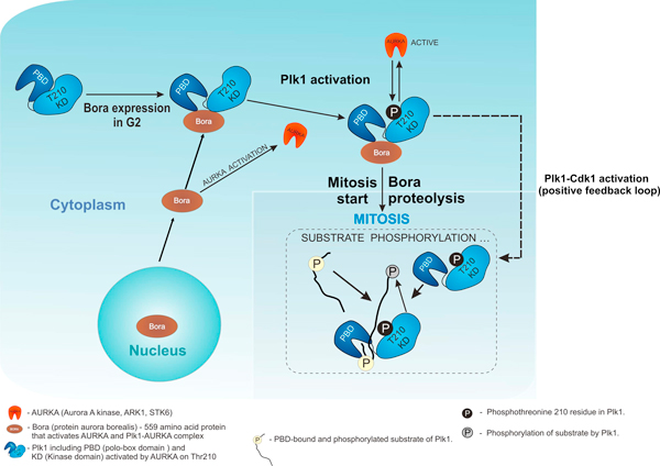
Interplay of Plk1, AURKA and BORA in cell cycle based on literature data: adapted from [43,46]and is based on references [29,38,43,44,46,57].
Similar articles
-
[Bioinformatic search of plant protein kinases, participating in microtubule protein phosphorylation and cell division regulation].Tsitol Genet. 2009 May-Jun;43(3):63-79. Tsitol Genet. 2009. PMID: 19938639 Russian.
-
[MAST2-like protein kinase from grape vine Vitis vinifera: cloning of catalytic domain cDNA].Tsitol Genet. 2010 Jul-Aug;44(4):41-7. Tsitol Genet. 2010. PMID: 20722285 Russian.
-
[Bioinformatic search for plant homologs of Ste20-like serine/threonine protein kinases].Tsitol Genet. 2009 Nov-Dec;43(6):68-77. Tsitol Genet. 2009. PMID: 20458979 Russian.
-
The Aurora kinases: role in cell transformation and tumorigenesis.Cancer Metastasis Rev. 2003 Dec;22(4):451-64. doi: 10.1023/a:1023789416385. Cancer Metastasis Rev. 2003. PMID: 12884918 Review.
-
TTBK2: a tau protein kinase beyond tau phosphorylation.Biomed Res Int. 2015;2015:575170. doi: 10.1155/2015/575170. Epub 2015 Apr 9. Biomed Res Int. 2015. PMID: 25950000 Free PMC article. Review.
Cited by
-
Understanding the Polo Kinase machine.Oncogene. 2015 Sep 10;34(37):4799-807. doi: 10.1038/onc.2014.451. Epub 2015 Jan 26. Oncogene. 2015. PMID: 25619835 Review.
-
Hyperosmolarity-induced suppression of group B1 Raf-like protein kinases modulates drought-growth trade-off in Arabidopsis.Proc Natl Acad Sci U S A. 2024 Dec 24;121(52):e2419204121. doi: 10.1073/pnas.2419204121. Epub 2024 Dec 19. Proc Natl Acad Sci U S A. 2024. PMID: 39700143 Free PMC article.
-
Regulation of cell cycle in plant gametes: when is the right time to divide?Development. 2025 Jan 15;152(2):dev204217. doi: 10.1242/dev.204217. Epub 2025 Jan 20. Development. 2025. PMID: 39831611 Free PMC article. Review.
-
MAST-like protein kinase IREH1 from Arabidopsis thaliana co-localizes with the centrosome when expressed in animal cells.Planta. 2017 Nov;246(5):959-969. doi: 10.1007/s00425-017-2742-4. Epub 2017 Jul 17. Planta. 2017. PMID: 28717875
-
Polo-like kinases: structural variations lead to multiple functions.Nat Rev Mol Cell Biol. 2014 Jul;15(7):433-52. doi: 10.1038/nrm3819. Nat Rev Mol Cell Biol. 2014. PMID: 24954208 Review.
References
-
- Amos LA. In: Nature Encyclopedia of Life Sciences. Zheng Y, Tickle C, Jansson R, Kehrer-Sawatzki H, Cooper DN, Melino G, Delves P, Battista J, Levitan I, Roberts K, Bynum WF, Harper H, editor. Chichester, John Wiley & Sons, Ltd; 2004. Tubulin and microtubules; pp. 1–12.http://www2.mrc-lmb.cam.ac.uk/archive/papers/2004-029.pdf
-
- McKean PG, Vaughan S, Gull K. The extended tubulin superfamily. J Cell Sci. 2001;114:2723–2733. - PubMed
-
- Demchuk ON, Blume YaB. Phylogenetic tree of bacterial and eukaryotic FtsZ-proteins based on the homology of their primary sequences. Tsitol Genet. 2005;39(4):3–12. - PubMed
Publication types
MeSH terms
Substances
LinkOut - more resources
Full Text Sources
Miscellaneous
