

Refinement of pig retroperfusion technique: Global retroperfusion with ligation of the azygos connection preserves hemodynamic function in an acute infarction model in pigs (Sus scrofa domestica)
- PMID: 20158947
- PMCID: PMC2826083
Refinement of pig retroperfusion technique: Global retroperfusion with ligation of the azygos connection preserves hemodynamic function in an acute infarction model in pigs (Sus scrofa domestica)
Abstract
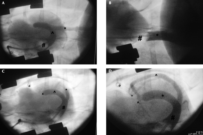
In ischemic hearts, venous retroperfusion is a potential myocardial revascularization strategy. This study aimed to refine the technical and functional aspects of a pig model of acute myocardial infarction and retroperfusion with respect to the azygos connection. Global retroperfusion after ligation of the ramus interventricularis paraconalis (equivalent to the left anterior descending artery in humans) was performed in 16 Landrace pigs (Sus scrofa domestica). Coronary sinus perfusion was performed in 8 pigs (P+) but not in the other 8 (P-), and the azygos vein was ligated (L+) 4 of the 8 pigs in each of these groups but left open (L-) in the remaining animals. Hemodynamic performance (for example, cardiac output, stroke volume) was significantly better in P+L+ pigs that underwent coronary sinus perfusion with ligation of the azygos vein compared with all other animals. In addition, troponin I release was significant lower in P+L+ pigs (1.7 +/- 1.3 ng/mL) than in P-L- (5.47 +/- 2.1 ng/mL), P-L+ (6.63 +/- 2.4 ng/mL), and P+L- (4.81 +/- 2.3 ng/mL) pigs. Effective retrograde flow and thus hemodynamic stability was achieved by ligation of the azygos vein. Therefore, experiments focusing on global retroperfusion will benefit from effective inhibition of the blood flow through the azygos vein.
Figures


References
-
- Agnew NM, Pennefather SH, Russell GN. 2002. Isoflurane and coronary heart disease. Anaesthesia 57:338–347 - PubMed
-
- Aldea GS, Zhang X, Rivers S, Shemin RJ. 1996. Salvage of ischemic myocardium with simplified and even delayed coronary sinus retroperfusion. Ann Thorac Surg 62:9–15 - PubMed
-
- Arealis EG, Volder JGR, Kolff WJ. 1973. Arterialization of the coronary vein coming from an ischemic area. Chest 63:462–463 - PubMed
-
- Beddard F. 1907. The azygos veins in the Mammalia. Proc Zool Soc Lond 2:10–20
-
- Bein B, Worthmann F, Tonner PH, Paris A, Steinfath M, Hedderich J, Scholz J. 2004. Comparison of esophageal Doppler, pulse contour analysis, and real-time pulmonary artery thermodilution for the continuous measurement of cardiac output. J Cardiothorac Vasc Anesth 18:185–189 - PubMed
Publication types
MeSH terms
Substances
LinkOut - more resources
Full Text Sources
Medical