

Topical and intravenous pilocarpine stimulated accommodation in anesthetized rhesus monkeys
- PMID: 20159011
- PMCID: PMC2909005
- DOI: 10.1016/j.exer.2010.02.005
Topical and intravenous pilocarpine stimulated accommodation in anesthetized rhesus monkeys
Abstract
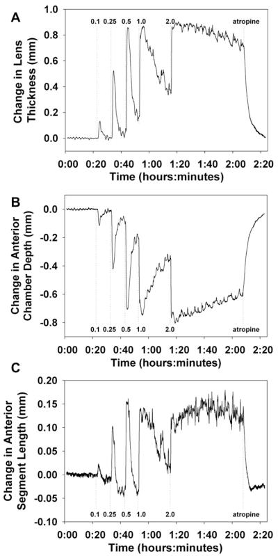
Many studies have used pilocarpine to stimulate accommodation in both humans and monkeys. However, the concentrations of pilocarpine used and the methods of administration vary. In this study, three different methods of pilocarpine administration are evaluated for their effectiveness in stimulating accommodation in rhesus monkeys. Experiments were performed in 17 iridectomized, anesthetized rhesus monkeys aged 4-16 years. Maximum accommodation was stimulated in all these monkeys with a 2% pilocarpine solution maintained on the cornea for at least 30 min in a specially designed perfusion lens. In subsequent topical pilocarpine experiments, baseline refraction was measured with a Hartinger coincidence refractometer and then while the monkeys were upright and facing forward, commercially available pilocarpine (2, 4, or 6%) was applied topically to the cornea as 2 or 4 drops in two applications or 6 drops in three applications over a five minute period with the eyelids closed between applications. Alternatively, while supine, 10-12 drops of pilocarpine were maintained on the cornea in a scleral cup for 5 min. Refraction measurements were begun 5 min after the second application of pilocarpine and continued for at least 30 min after initial administration until no further change in refraction occurred. In intravenous experiments, pilocarpine was given either as boluses ranging from 0.1mg/kg to 2mg/kg or boluses followed by a constant infusion at rates between 3.06 mg/kg/h and 11.6 mg/kg/h. Constant 2% pilocarpine solution on the eye in the perfusion lens produced 10.88+/-2.73 D (mean+/-SD) of accommodation. Topically applied pilocarpine produced 3.81 D+/-2.41, 5.49 D+/-4.08, and 5.55 D+/-3.27 using 2%, 4%, and 6% solutions respectively. When expressed as a percentage of the accommodative response amplitude obtained in the same monkey with constant 2% pilocarpine solution on the eye, the responses were 34.7% for 2% pilocarpine, 48.4% for 4% pilocarpine, and 44.6% for 6% pilocarpine. Topical 4% and 6% pilocarpine achieved similar, variable accommodative responses, but neither achieved maximum accommodation. IV boluses of pilocarpine achieved near maximal levels of accommodation at least ten times faster than topical methods. Doses effective for producing maximum accommodation ranged from 0.25mg/kg to 1.0mg/kg. IV pilocarpine boluses caused an anterior movement of the anterior lens surface, a posterior movement of the posterior lens surface, and a slight net anterior movement of the entire lens. Considerable variability in response amplitude occurred and maximum accommodative amplitude was rarely achieved with topical application of a variety of concentrations of commercially available pilocarpine. Intravenous infusion of pilocarpine was a rapid and reliable method of producing a nearly maximal accommodative response and maintaining accommodation when desired.
(c) 2010 Elsevier Ltd. All rights reserved.
Figures


Similar articles
-
Comparison between carbachol iontophoresis and intravenous pilocarpine stimulated accommodation in anesthetized rhesus monkeys.Exp Eye Res. 2013 Oct;115:123-30. doi: 10.1016/j.exer.2013.06.028. Epub 2013 Jul 11. Exp Eye Res. 2013. PMID: 23850971
-
Autonomic drugs and the accommodative system in rhesus monkeys.Exp Eye Res. 2010 Jan;90(1):104-12. doi: 10.1016/j.exer.2009.09.015. Epub 2009 Sep 24. Exp Eye Res. 2010. PMID: 19782072 Free PMC article.
-
Lens diameter and thickness as a function of age and pharmacologically stimulated accommodation in rhesus monkeys.Exp Eye Res. 2008 May;86(5):746-52. doi: 10.1016/j.exer.2008.01.022. Epub 2008 Feb 8. Exp Eye Res. 2008. PMID: 18342856 Free PMC article.
-
Parasympathetic denervation of the ciliary muscle following retinal photocoagulation.Trans Am Ophthalmol Soc. 1990;88:513-53. Trans Am Ophthalmol Soc. 1990. PMID: 2095033 Free PMC article. Review.
-
Accommodating intraocular lenses: a critical review of present and future concepts.Graefes Arch Clin Exp Ophthalmol. 2007 Apr;245(4):473-89. doi: 10.1007/s00417-006-0391-6. Epub 2006 Aug 30. Graefes Arch Clin Exp Ophthalmol. 2007. PMID: 16944188 Review.
Cited by
-
Presbyopia: a new potential pharmacological treatment.Med Hypothesis Discov Innov Ophthalmol. 2012 Spring;1(1):3-5. Med Hypothesis Discov Innov Ophthalmol. 2012. PMID: 24600609 Free PMC article.
-
Intraocular accommodative movements in monkeys; relationship to presbyopia.Exp Eye Res. 2022 Sep;222:109029. doi: 10.1016/j.exer.2022.109029. Epub 2022 Mar 11. Exp Eye Res. 2022. PMID: 35283107 Free PMC article.
-
Influence of Pilocarpine Eyedrops on the Ocular Biometric Parameters and Intraocular Lens Power Calculation.J Ophthalmol. 2023 Jul 7;2023:7680659. doi: 10.1155/2023/7680659. eCollection 2023. J Ophthalmol. 2023. PMID: 37455794 Free PMC article.
-
Presbyopia Treatment With Eye Drops: An Eight Year Retrospective Study.Transl Vis Sci Technol. 2020 Jun 23;9(7):25. doi: 10.1167/tvst.9.7.25. eCollection 2020 Jun. Transl Vis Sci Technol. 2020. PMID: 32832231 Free PMC article.
-
Safety and Efficacy of AGN-190584 in Individuals With Presbyopia: The GEMINI 1 Phase 3 Randomized Clinical Trial.JAMA Ophthalmol. 2022 Apr 1;140(4):363-371. doi: 10.1001/jamaophthalmol.2022.0059. JAMA Ophthalmol. 2022. PMID: 35238902 Free PMC article. Clinical Trial.
References
-
- Asseff CF, Weisman RL, Podos SM, Becker B. Ocular penetration of pilocarpine in primates. Am. J. Ophthalmol. 1973;75:212–215. - PubMed
-
- Beers APA, van der Heijde GL. In vivo determination of the biomechanical properties of the component elements of the accommodative mechanism. Vision Res. 1994;34:2897–2905. - PubMed
-
- Bito LZ, DeRousseau CJ, Kaufman PL, Bito JW. Age-dependent loss of accommodative amplitude in rhesus monkeys: an animal model for presbyopia. Invest. Ophthalmol. Vis. Sci. 1982;23:23–31. - PubMed
-
- Bito LZ, Kaufman PL, DeRousseau CJ, Koretz J. Presbyopia: an animal model and experimental approaches for the study of the mechanism of accommodation and ocular aging. Eye. 1987;1(2):222–230. - PubMed
Publication types
MeSH terms
Substances
Grants and funding
LinkOut - more resources
Full Text Sources
