. 2010 May;44(1):43-54.
doi: 10.1016/j.mcn.2010.02.002. Epub 2010 Feb 14.

High content screening of cortical neurons identifies novel regulators of axon growth

Affiliations

High content screening of cortical neurons identifies novel regulators of axon growth

Murray G Blackmore et al. Mol Cell Neurosci. 2010 May.

Abstract

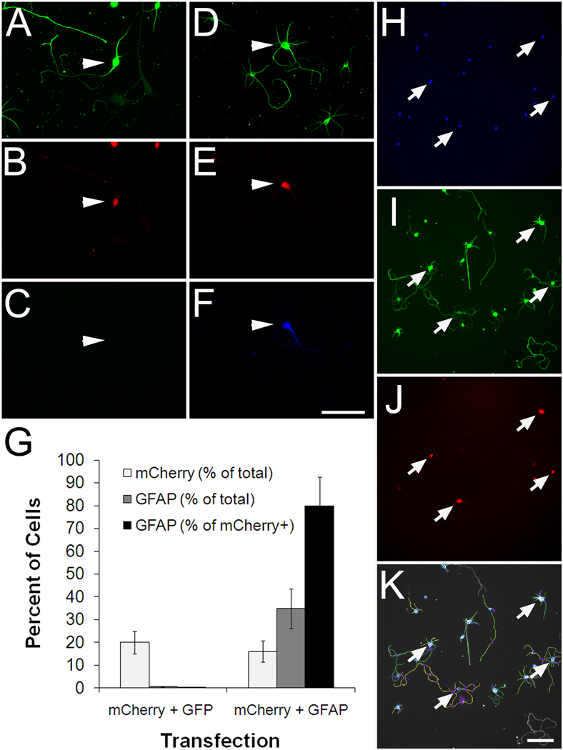
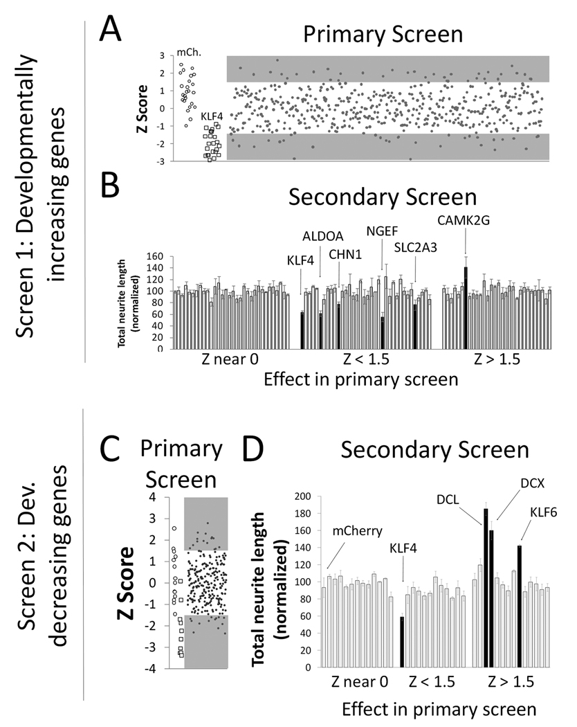
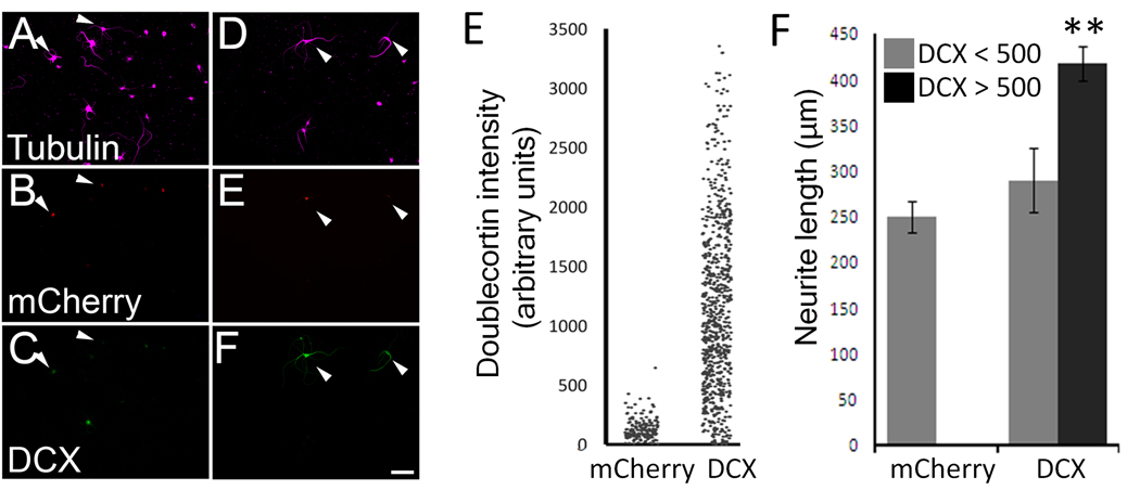
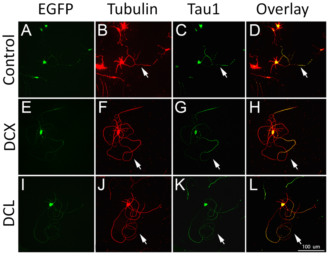
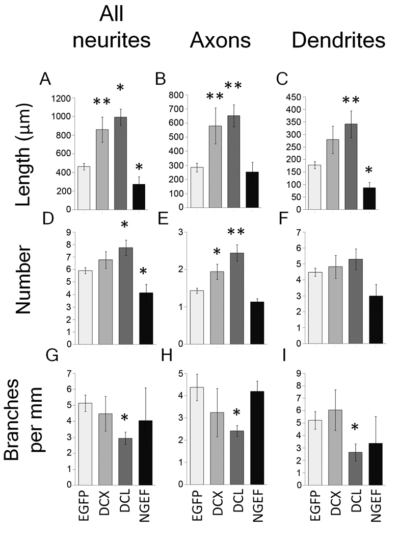
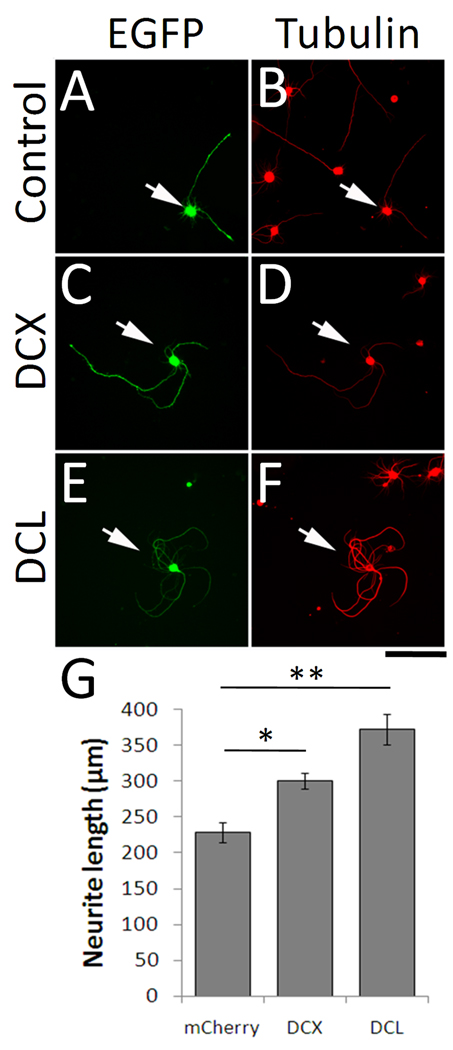
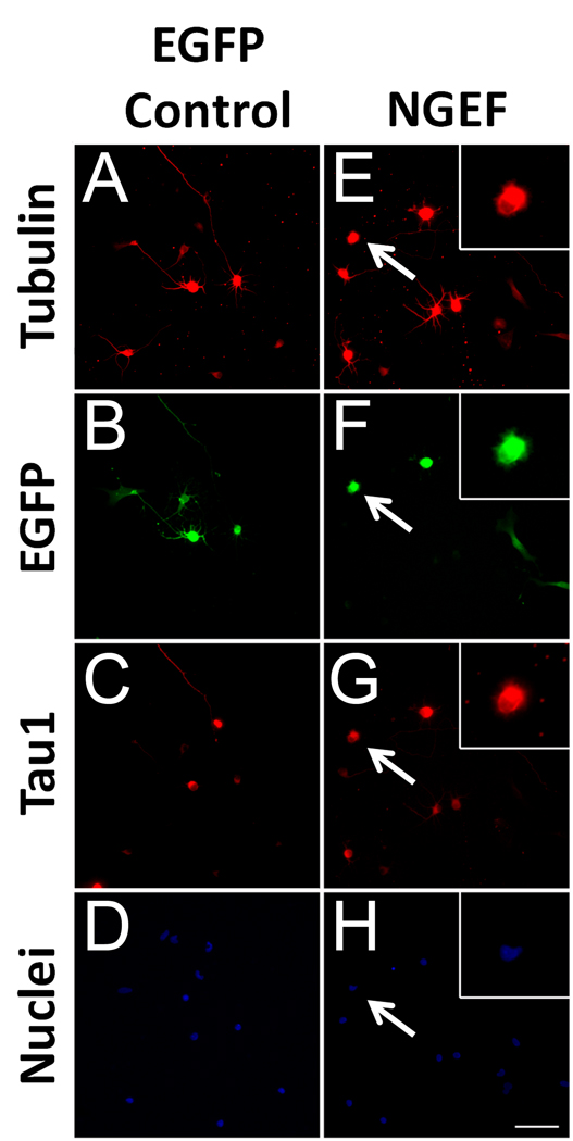
Neurons in the central nervous system lose their intrinsic capacity for axon regeneration as they mature, and it is widely hypothesized that changes in gene expression are responsible. Testing this hypothesis and identifying the relevant genes has been challenging because hundreds to thousands of genes are developmentally regulated in CNS neurons, but only a small subset are likely relevant to axon growth. Here we used automated high content analysis (HCA) methods to functionally test 743 plasmids encoding developmentally regulated genes in neurite outgrowth assays using postnatal cortical neurons. We identified both growth inhibitors (Ephexin, Aldolase A, Solute Carrier 2A3, and Chimerin), and growth enhancers (Doublecortin, Doublecortin-like, Kruppel-like Factor 6, and CaM-Kinase II gamma), some of which regulate established growth mechanisms like microtubule dynamics and small GTPase signaling. Interestingly, with only one exception the growth-suppressing genes were developmentally upregulated, and the growth-enhancing genes downregulated. These data provide important support for the hypothesis that developmental changes in gene expression control neurite outgrowth, and identify potential new gene targets to promote neurite outgrowth.


Figures

Figure 1
Figure 1. Cortical neurons transfected in 96-well format are identified by co-transfected mCherry reporter and efficiently traced by Cellomics instrumentation
(A–G) P1 cortical neurons were co-transfected with mCherry and either EGFP or GFAP. (A–C) Control neurons transfected with mCherry and EGFP express βIII tubulin (arrowhead, A) and mCherry (arrowhead, B), but do not express GFAP (arrowhead, C). (D–E) Neurons transfected with mCherry and GFAP express both mCherry (arrow, E) and GFAP (arrow, F). (G) Quantification shows that 80% of mCherry-positive neurons express GFAP after co-transfection. More than 300 neurons were quantified per condition. (H–K) P1 cortical neurons transfected with mCherry were cultured for 3 days prior to fixation, staining, and automated scanning (Kineticscan, Cellomics). Only cells identified as neurons by βIII tubulin staining (I), and successfully transfected with mCherry reporter (arrows, J) were included in subsequent analysis. (D) Digitized overlay shows effective tracing of cell processes. Scale bars, 100µm.
Figure 2
Figure 2. An overexpression screen of developmentally regulated genes
Cortical neurons (P1, A, B; P5, C,D) were co-transfected with test genes and mCherry, and cultured for 3 days. (A, C) In primary screens of both up-regulated (A) and down-regulated (C) genes, plasmids were tested in sets of 24 that included three controls (no plasmid, mCherry (open circles), and growth-suppressive KLF4 (open squares)). Average neurite length was calculated from at least 100 transfected neurons, and a Z-scores was calculated for each gene based on the average and standard deviation across all test genes in the set (see Methods). (A, C) Each point represents the Z score from a plasmid in one experiment. , and shaded areas indicate plasmids selected for re-screening (Z scores > or < 1.5). (B,D) In secondary screens, genes were tested in sets of 24 that included mCherry (leftmost bar) and eight genes with no effect in the primary screen (Z near 0; see Methods). Neurite length was normalized to the average of the control genes. Bars represent the average normalized length across three replicate experiments. Black bars represent genes that differed significantly from controls (P < 0.05, ANOVA with post-hoc Dunnett’s).
Figure 3
Figure 3. DCX overexpression correlates with longer neurites
P5 cortical neurons were transfected with mCherry ± doublecortin. (A–F). Control neurons express mCherry (arrows, B) and are faintly immunoreactive for DCX. (C). Neurons co-transfected with mCherry and doublecortin (D–F) express mCherry reporter (E) and are brightly immunoreactive for DCX (F). (G) Each dot represents the average DCX immunofluorescence intensity of a single transfected (mCherry+) neuron. Nearly 100% of control neurons had an average intensity below 500 arbitrary units, while 87% of DCX-transfected neurons had an average intensity above 500. (H) Average neurite length of transfected neurons (βIII tubulin+, mCherry+). Neurons that were brightly immunofluorescent for DCX extended longer neurites that those with dimmer immunofluorescence (P<0.01, paired t-test, n>300 in each treatment). Scale bar is 50µm.
Figure 4
Figure 4. Overexpression of DCX and DCL increase axon length in postnatal cortical neurons
P5 cortical neurons were transfected with EGFP (A–D), or EGFP plus DCX (E–H) or DCL (I–L). After three days, neurons were immunostained for βIII tubulin (B,F,J) and Tau1 (C,G,K). Arrows identify axons from transfected (EGFP+) neurons. Compared to controls, neurons transfected with DCX or DCL extend long axons with looped trajectories (arrows, H, L). Scale bar is 100µm.
Figure 5
Figure 5. DCX, DCL, and NGEF affect length, number, and branching of axons and dendrites
P5 cortical neurons were transfected with EGFP (control), or co-transfected with EGFP and DCX, DCL, or NGEF. Images of EGFP+ neurons were acquired and traced using Neurolucida. All EGFP+ neurons with neurites were included in A, D and G. For other graphs, only polarized neurons with clear tau1-positive processes were included. (A–C) NGEF decreased and DCX and DCL increased average neurite length. DCX and DCL significantly increased the length of axons, and DCL significantly increased dendrite length. (D–F) NGEF decreased and DCL increased the average number of neurites, whereas DCX and DCL significantly increased the average number of axons (E), but not the number of dendrites (F). (G–H) DCL significantly decreased the amount of branching for both axon and dendrites. N ≥ 40. *p<0.05, **p<0.01 ANOVA with post-hoc Dunnett’s.
Figure 6
Figure 6. DCX is regulated in developing cortex and in RGCs
(A–D) Horizontal cortical sections and transverse spinal cord sections from P1 and adult animals were immunostained for DCX (green). DCX Immunoreactivity is bright throughout P1 cortex (A) and in the CST in P1 spinal cord (arrow, C), but is very dim in adult cortex (B) and spinal cord (D). (E–J) Retinas (E–H) and optic nerves (I,J) were cryosectioned and stained for DCX. DCX immunoreactivity (green) is abundant in the retinal ganglion cell layer at E15 and P1 (asterisk, E,F) but is downregulated by P9 (G) and undetectable in the adult (H). In the optic nerve, DCX is readily detectable at P1 (I), but undetectable in the adult (J). Scale bars are 100µm (A–D, I,J) and 50 µm (E–H).
Figure 7
Figure 7. Overexpression of DCX or DCL increases neurite length in postnatal RGCs
Purified P4 RGCs were transfected with EGFP (control) or EGFP plus DCX or DCL, cultured for 2 days, and stained for tubulin. Transfected RGCs were identified by EGFP (arrows). Compared to control (A,B), RGCs transfected with DCX (C,D) or DCL (E,F) showed increased neurite outgrowth and curved/looped neurite trajectories. (G) Total neurite length was significantly increased in neurons transfected with DCX or DCL (*p<0.05, **p<0.01, ANOVA with post-hoc Dunnet’s). N= 3 experiments, with > 100 transfected neurons/treatment. Error bars show SEM. Scale bar, 100µm.
Figure 8
Figure 8. NGEF suppresses neurite growth in postnatal cortical neurons
P1 cortical neurons were co-transfected with EGFP (control, A–D) or NGEF plus EGFP (NGEF, E–H) and cultured for 3 days. Control neurons (A–D) extend long processes, some of which are identified as axons by Tau1 immunoreactivity (C). Neurons transfected with NGEF (E–H, arrows) often extended no neurites but appeared viable with large, uncondensed nuclei and abundant lamellar protrusions (insets, E–H). Scale bar is 50µm.

Similar articles

Cited by

References

    1. Arlotta P, Molyneaux BJ, Chen J, Inoue J, Kominami R, Macklis JD. Neuronal subtype-specific genes that control corticospinal motor neuron development in vivo. Neuron. 2005;45:207–221. - PubMed
    1. Barres BA, Silverstein BE, Corey DP, Chun LL. Immunological, morphological, and electrophysiological variation among retinal ganglion cells purified by panning. Neuron. 1988;1:791–803. - PubMed
    1. Benson MD, Romero MI, Lush ME, Lu QR, Henkemeyer M, Parada LF. Ephrin-B3 is a myelin-based inhibitor of neurite outgrowth. Proc Natl Acad Sci U S A. 2005;102:10694–10699. - PMC - PubMed
    1. Bielas SL, Serneo FF, Chechlacz M, Deerinck TJ, Perkins GA, Allen PB, Ellisman MH, Gleeson JG. Spinophilin facilitates dephosphorylation of doublecortin by PP1 to mediate microtubule bundling at the axonal wrist. Cell. 2007;129:579–591. - PMC - PubMed
    1. Blackmore M, Letourneau PC. Changes within maturing neurons limit axonal regeneration in the developing spinal cord. J Neurobiol. 2006;66:348–360. - PubMed

Publication types

MeSH terms