. 2010 Feb 11;65(3):358-72.
doi: 10.1016/j.neuron.2010.01.007.

Regulation of Rap2A by the ubiquitin ligase Nedd4-1 controls neurite development

Affiliations

Regulation of Rap2A by the ubiquitin ligase Nedd4-1 controls neurite development

Hiroshi Kawabe et al. Neuron. .

Abstract

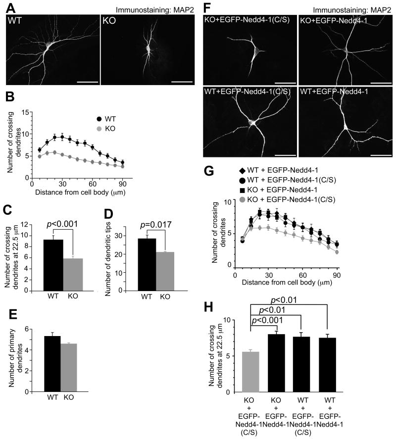
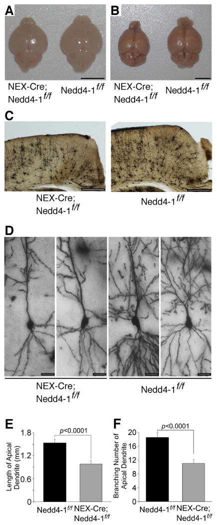
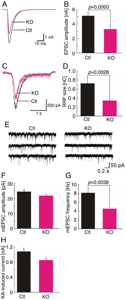
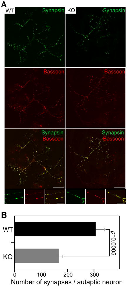
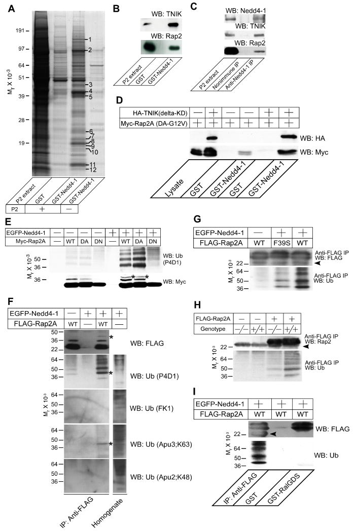
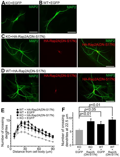
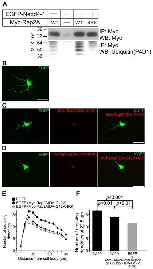
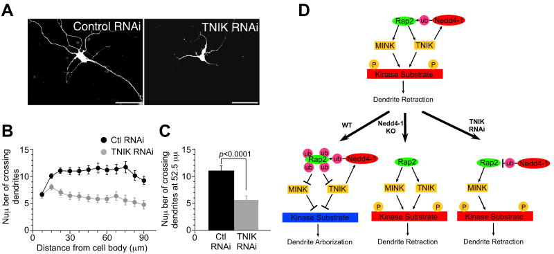
Nedd4-1 is a "neuronal precursor cell expressed and developmentally downregulated protein" and among the most abundant E3 ubiquitin ligases in mammalian neurons. In analyses of conventional and conditional Nedd4-1-deficient mice, we found that Nedd4-1 plays a critical role in dendrite formation. Nedd4-1, the serine/threonine kinase TNIK, and Rap2A form a complex that controls Nedd4-1-mediated ubiquitination of Rap2A. Ubiquitination by Nedd4-1 inhibits Rap2A function, which reduces the activity of Rap2 effector kinases of the TNIK family and promotes dendrite growth. We conclude that a Nedd4-1/Rap2A/TNIK signaling pathway regulates neurite growth and arborization in mammalian neurons.


Figures

Figure 1
Figure 1. Impaired Dendrite Development in Nedd4-1-KO Neurons
(A) KO neurons show impaired development of dendrites. Autaptic neurons prepared from cortices of WT and KO embryos were fixed at DIV10 and dendrites were immunostained using an anti-MAP2 antibody. Scale bars, 50 μm. (B) Sholl analysis of WT and KO neurons. Black circles, WT neurons (n=25); gray circles, KO neurons (n=38). (C) Numbers of crossing dendrites at 22.5 μm from the cell body in WT and KO neurons. (D) Numbers of dendritic tips in WT and KO neurons. (E) Numbers of primary dendrites emerging from WT and KO neurons. Black bars, WT neurons (n=25 for C, n=26 for D and E); gray bars, KO neurons (n=38 for C, n=39 for D and E). Student's t-test revealed significant differences in (C) (p<0.001) and (D) (p=0.017), but not in (E) (p=0.17). (F) Anti-MAP2 immunostaining of autaptic KO (top two panels) and WT (bottom two panels) neurons overexpressing EGFP fused to the catalytically inactive mutant of Nedd4-1 (C/S) as a negative control (left two panels) or full length EGFP-Nedd4-1 (right two panels). Scale bars, 50 μm. (G) Sholl analysis of autaptic WT or KO neurons overexpressing EGFP-Nedd4-1 (C/S) or EGFP-Nedd4-1. Black diamonds, WT neurons overexpressing EGFP-Nedd4-1 (n=42); black circles, WT neurons overexpressing EGFP-Nedd4-1 (C/S) (n=40); black rectangles, KO neurons overexpressing EGFP-Nedd4-1 (n=60); gray circles, KO neurons overexpressing EGFP-Nedd4-1(C/S) (n=68). (H) Numbers of crossing dendrites at 22.5 μm from the cell body in WT and KO neurons expressing EGFP-Nedd4-1(C/S) or EGFP-Nedd4-1. nonparametric ANOVA test revealed a significant effect of interaction between the genotype and expression of EGFP-Nedd4-1s (p=0.0001). Dunn's multiple comparisons test revealed significant differences between KO neurons expressing EGFP-Nedd4-1(C/S) and EGFP-Nedd4-1 (p<0.001) and between EGFP-Nedd4-1(C/S) expressing KO and WT neurons (p<0.01) but not between WT neurons expressing EGFP-Nedd4-1(C/S) and EGFP-Nedd4-1 (p >0.05). See also Figures S1-S4, and Tables S1 and S2.
Figure 2
Figure 2. Effect of Nedd4-1 Loss on Dendrite Development In Vivo
(A and B) Impaired development of cerebra of NEX-Cre;Nedd4-1flox/flox mice (left) as compared to control Nedd4-1flox/flox mice (right) at immature (P5; A) and mature (8 months; B) stages. Note that the sizes of cerebella are indistinguishable between NEX-Cre;Nedd4-1flox/flox and Nedd4-1flox/flox mice. Scale bars, 5 mm. (C) Overviews of Golgi stained samples of NEX-Cre;Nedd4-1flox/flox and Nedd4-1flox/flox cerebra. Sections were from two months old littermates. Scale bars, 0.5 mm. (D) Hippocampal CA1 neurons in NEX-Cre;Nedd4-1flox/flox (left two panels) and Nedd4-1flox/flox (right two panels) mice. Tops of each picture are the apical side. Pictures were taken from three months old littermates. Scale bars, 20 μm. (E and F) Total lengths (E) and branching numbers (F) of apical dendrites of hippocampal CA1 neurons in NEX-Cre;Nedd4-1flox/flox and Nedd4-1flox/flox mice. Similar numbers of neurons were used for each genotype at the age of two months (NEX-Cre;Nedd4-1flox/flox, n=3; Nedd4-1flox/flox, n=3), three months (NEX-Cre;Nedd4-1flox/flox, n=16; Nedd4-1flox/flox, n=13), and eight months (NEX-Cre;Nedd4-1flox/flox, n=9; Nedd4-1flox/flox, n=9). Student's t-test revealed significant differences in both (E) and (F) (n>24, p<0.0001). See also Figures S1, S2, and Table S1.
Figure 3
Figure 3. Reduced Synaptic Transmission in Nedd4-1-KO Neurons
(A) Sample traces of evoked EPSCs from control (Ctl) and KO neurons. (B) Averaged evoked EPSC amplitudes of control (Ctl) (n=70) and KO (n=82) neurons. Student's t-test, p=0.0003. (C) Sample traces of postsynaptic currents evoked by the application of hypertonic sucrose solution from control (Ctl) and KO neurons. (D) Mean readily releasable vesicle pool sizes as estimated by the charge integral measured after release induced by application of 0.5 M sucrose solution in control (Ctl) (n=58) and KO (n=61) neurons. Student's t-test, p=0.0028. (E) Sample traces of miniature EPSCs from control (Ctl) and KO neurons. (F) Averaged miniature EPSC amplitudes (Ctl, n=32; KO, n=40). Student's t-test, p>0.1. (G) Averaged miniature EPSC frequencies (Ctl, n=32; KO, n=40). Student's t-test, p=0.0038. (H) Averaged kainic acid-induced currents in control (Ctl, n=29) and KO neurons (n=32). Student's t-test, p>0.1. See also Figures S3 and S4 and Table S3.
Figure 4
Figure 4. Reduced Synapse Numbers in Nedd4-1-KO Neurons
(A) Immunocytochemical analysis of synapses in autaptic WT and KO neurons using anti-Synapsin and anti-Bassoon antibodies. Cortical neurons were prepared from embryos at E13 and fixed at DIV10. Note the smaller size of the KO neuron. Scale bars, 50 μm (low magnification) and 20 μm (high magnification insets). (B) Quantification of numbers of Synapsin and Bassoon double positive synaptic puncta in individual autaptic KO (n=37) and WT neurons (n=42). Student's t-test, p=0.0005. See also Figures S3 and S4 and Table S3.
Figure 5
Figure 5. Rap2 is a Target of Nedd4-1
(A) Affinity purification of TNIK as a binding partner of Nedd4-1. 40 μg of GST or GST Nedd4-1 (residues 217-549) were immobilized on glutathione Sepharose beads and a Triton X-100 extract of rat brain synaptosomes (P2 extract, +) or buffer (-) were applied. After washing the beads, bound proteins were eluted with 1 M NaCl. Protein bands that appeared to be enriched in the eluate from the GST Nedd4-1 column were analyzed by mass spectrometry. Protein identification was successful for the twelve marked bands. The results of the mass spectrometric analysis are given in Table S4. (B) Complex formation of Rap2 with TNIK and GST-Nedd4-1. Samples eluted from the Nedd4-1 beads (see A) were blotted for TNIK (top) and Rap2 (bottom). Neither antibody cross-reacted with samples purified on GST beads that were used as a negative control. (C) Complex formation of endogenous Rap2, TNIK, and Nedd4-1. Nedd4-1 was immunoprecipitated using a rabbit polyclonal anti-Nedd4-1 antibody from mouse brain membranes after treatment with a thiol-cleavable chemical crosslinker. Precipitates were boiled in Laemmli buffer with 50 mM DTT, loaded to SDS-PAGE gels, and analyzed by Western blotting using anti-Nedd4-1, anti-TNIK, or anti-Rap2 antibodies. Note that TNIK and Rap2 were coimmunoprecipitated with Nedd4-1 but not with the negative control IgG. (D) TNIK dependent interaction of Rap2A with Nedd4-1. GST or GST-Nedd4-1 (residues 217-549) were immobilized on glutathione Sepharose beads and recombinant Myc-Rap2A(DA-G12V) was loaded in the presence (+) or absence (-) of HA-TNIK (delta-KD). Interaction was detected by Western blotting using anti-HA or anti-Myc antibodies. (E) Ubiquitination of Rap2A by Nedd4-1. EGFP-Nedd4-1 (+) or EGFP alone (-) were coexpressed with WT, dominant active (DA), or dominant negative (DN) mutants of Myc-tagged Rap2A. Myc-Rap2A was immunoprecipitated using an anti-Myc antibody. Immunoprecipitates were blotted for Myc (lower panel) and ubiquitin (upper panel). (F) Mono- and di-ubiquitination of Rap2A by Nedd4-1. FLAG-Rap2A was immunoprecipitated from HEK cells expressing FLAG-Rap2A with or without EGFP-Nedd4-1. FLAG-Rap2A was eluted from anti-FLAG antibody coupled beads with 3×FLAG peptides and immunoblotted using four different monoclonal mouse or rabbit anti-ubiquitin antibodies, P4D1, FK1, Apu3, and Apu2. P4D1 recognizes both poly- and monoubiquitin conjugated proteins while FK1 recognizes only polyubiquitin conjugated ones. Apu3 and Apu2 recognize K63-linked and K48-linked polyubiquitin chains, respectively. The lysate of HEK cells overexpressing EGFP-Nedd4-1 was also blotted using the four anti-ubiquitin antibodies in order to show that the antibody titers are comparable. (G) Loss of TNIK interaction in the F39S point mutant of Rap2 reduces the ubiqutination of Rap2 by Nedd4-1. WT or F39S point mutant FLAG-Rap2A were overexpressed in HEK cells together with EGFP (-) or EGFP-Nedd4-1 (+). Efficiencies of immunoprecipitation of FLAG-Rap2A were comparable as seen in the Western blot using the anti-FLAG antibody (upper panel). FLAG-Rap2A(F39S) showed clearly weaker intensities of ladder patterns than FLAG-Rap2A(WT) in the blot using the anti-ubiquitin antibody (lower panel). (H) Ubiquitination of Rap2A by endogenous Nedd4-1 and ubiquitin. WT (+/+) and Nedd4-1-KO (-/-) MEFs were transfected with a mammalian expression vector encoding FLAG-Rap2A. Proteins from lysed cells were immunoprecipiated with an anti-FLAG antibody and blotted for Rap2A with an anti-Rap2 antibody (upper panel) and an anti-ubiquitin antibody (lower panel). (I) Ubiquitination of Rap2A blocks the Rap2A function. FLAG-Rap2A(WT) expressed in HEK cells together with EGFP-Nedd4-1 was precipitated using anti-FLAG antibodies (first lane) or GST-RalGDS-coupled beads (third lane), and blotted for FLAG (upper panel) or ubiquitin (lower panel). Note that ubiquitinated FLAG-Rap2A was efficiently precipitated only with the anti-FLAG antibody while total amounts of precipitated FLAG-Rap2A were comparable between lanes 1 and 3. Arrowheads in (G-I) indicate the light chain of the anti-FLAG IgG used for immunoprecipitation. All results shown are representative of two to three independent experiments. Asterisks in (E) and (F) indicate bands with a molecular weight corresponding to Myc-Rap2A or FLAG-Rap2A conjugated with two ubiquitin moieties. See also Figures S1, S5, and S6, and Table S4.
Figure 6
Figure 6. Overexpression of a Dominant Negative Mutant of Rap2A Rescues the Dendritogenesis Defect in Nedd4-1-KO Neurons
(A-D) Anti-MAP2 immunostaining of KO or WT neurons overexpressing EGFP (A and B), and anti-MAP2 and anti-HA immunostaining of KO or WT neurons overexpressing dominant negative HA-Rap2A(DN-S17N) (C and D). HA-Rap2A(DN-S17N) overexpressing neurons showed longer and more complex dendrites. This effect was more evident in KO neurons. Scale bars, 50 μm. (E) Sholl analysis of KO neurons overexpressing EGFP (gray circles, n=68) or HA-Rap2ADN (black rectangles, n=22), and of WT neurons overexpressing EGFP (black circles, n=20) or HA-Rap2ADN (black diamonds, n=12). (F) Numbers of crossing dendrites at 22.5 μm from the cell body in WT and KO neurons overexpressing EGFP or HA-Rap2ADN. Nonparametric ANOVA test revealed a significant effect of interaction between the genotype and expression of HA-Rap2A(DN-S17N) (p<0.0001). Dunn's multiple comparisons test revealed significant differences between KO neurons expressing EGFP and HA-Rap2A(DN-S17N) (p<0.01), and between KO and WT neurons expressing EGFP (p<0.05), but not between WT neurons expressing EGFP and HA-Rap2A(DN-S17N) (p >0.05). See also Figures S4 and S6-S8, and Table S2.
Figure 7
Figure 7. Perturbation of Ubiquitination of Rap2A Enhances Rap2A Function in Cultured Neurons
(A) HEK cells were transfected with Myc-tagged WT or 4RK mutant variants of Rap2A, with or without EGFP-Nedd4-1. Myc-Rap2A was immunoprecipitated with anti-Myc antibodies, and ubiquitination was detected with the anti-ubiquitin P4D1 monoclonal antibody (Jura et al., 2006). (B-D) Overexpression of Myc-Rap2A(DA-G12V;4RK) reveals a gain-of-function phenotype. Myc-Rap2A (DA-G12V) and EGFP coexpressing neurons shows impaired development of neurites as compared to EGFP expressing neurons (B, C). This effect of Rap2A was more pronounced when Myc-Rap2A(DA-G12V;4RK) was coexpressed with EGFP (D). Scale bar, 50 μm. (E) Sholl analysis of Rap2A overexpressing neurons. Black rectangles, EGFP- expressing neurons (n=80); black circles, EGFP- and Myc-Rap2A(DA-G12V) coexpressing neurons (n=109); gray circles, EGFP- and Myc-Rap2A(DA-G12V;4RK) coexpressing neurons (n=109). (F) Numbers of crossing dendrites at 22.5 μm from the cell body in EGFP-, EGFP- and Myc-Rap2A(G12V)-, or EGFP- and Myc-Rap2A(DA-G12V;4RK)-over expressing neurons. Nonparametric ANOVA test revealed a significant effect of expression of Myc-Rap2 mutants (p<0.0001). Dunn's multiple comparisons test revealed significant differences between groups linked with black lines. See also Figures S6-S8 and Table S2.
Figure 8
Figure 8. RNAi Silencing of Endogenous TNIK Phenocopies the Effect of Nedd4-1 Loss in Neurons
(A) WT mouse neurons transfected with the vector encoding an shRNA expression cassette for TNIK knock-down showed impaired development of neurites (right panel) while control shRNA expressing neurons showed normal morphology (left panel). Scale bar; 50 μm. (B) Sholl analysis of TNIK knock-down neurons. Black circles, control knockdown neurons (n=37); gray circles, TNIK-knock-down neurons (n=26). (C) Numbers of crossing dendrites at 52.5 mm from the cell body in control and TNIK-knock-down neurons. Student's t-test; p<0.0001. (D) Model of Nedd4-1 function in the regulation of neurite development. Given that only a fraction of total Rap2 is ubiquitinated, the depicted signaling pathway must be restricted (i.e. subcellularly compartmentalized) to growing neurite compartments by an as yet unknown mechanism. See Discussion for details. See also Figures S6-S8 and Table S2.

Comment in

  • Nedd4 branches out.
    DiAntonio A. DiAntonio A. Neuron. 2010 Feb 11;65(3):293-4. doi: 10.1016/j.neuron.2010.01.028. Neuron. 2010. PMID: 20159442 Free PMC article.

References

    1. Arevalo JC, Waite J, Rajagopal R, Beyna M, Chen ZY, Lee FS, Chao MV. Cell survival through Trk neurotrophin receptors is differentially regulated by ubiquitination. Neuron. 2006;50:549–559. - PubMed
    1. Barnes AP, Lilley BN, Pan YA, Plummer LJ, Powell AW, Raines AN, Sanes JR, Polleux F. LKB1 and SAD kinases define a pathway required for the polarization of cortical neurons. Cell. 2007;129:549–563. - PubMed
    1. Bekkers JM, Stevens CF. Excitatory and inhibitory autaptic currents in isolated hippocampal neurons maintained in cell culture. Proc Natl Acad Sci U S A. 1991;88:7834–7838. - PMC - PubMed
    1. Bloom AJ, Miller BR, Sanes JR, DiAntonio A. The requirement for Phr1 in CNS axon tract formation reveals the corticostriatal boundary as a choice point for cortical axons. Genes Dev. 2007;21:2593–2606. - PMC - PubMed
    1. Cao XR, Lill NL, Boase N, Shi PP, Croucher DR, Shan H, Qu J, Sweezer EM, Place T, Kirby PA, et al. Nedd4 controls animal growth by regulating IGF-1 signaling. Sci Signal. 2008;1:ra5. - PMC - PubMed

Publication types

MeSH terms