

Review
doi: 10.1016/j.molcel.2010.01.025.
The BCL-2 family reunion
Affiliations
- PMID: 20159550
- PMCID: PMC3222298
- DOI: 10.1016/j.molcel.2010.01.025
Item in Clipboard
Review
The BCL-2 family reunion
Mol Cell.
.
Abstract
B cell CLL/lymphoma-2 (BCL-2) and its relatives comprise the BCL-2 family of proteins, which were originally characterized with respect to their roles in controlling outer mitochondrial membrane integrity and apoptosis. Current observations expand BCL-2 family function to include numerous cellular pathways. Here we will discuss the mechanisms and functions of the BCL-2 family in the context of these pathways, highlighting the complex integration and regulation of the BCL-2 family in cell fate decisions.
Figures
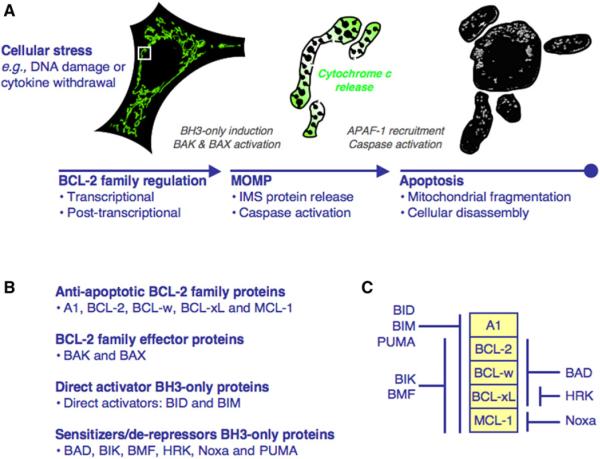
(A) Cellular stress causes transcriptional and post-transcriptional regulation of the BCL-2 family to promote MOMP.MOMP is induced by interactions between the BH3-only and effector proteins and leads to cytochrome c release, APAF-1 recruitment, and caspase activation. At the time of MOMP (middle), the intact mitochondrial network (green, left) undergoes fragmentation (gray, right), and soon after the cell is disassembled. The mitochondria in the middle are enlarged from the white box. (B) The BCL-2 family is divided into antiapoptotic, effector, and direct activator/sensitizer/derepressor BH3-only proteins. (C) The antiapoptotic BCL-2 protein binding profiles for the BH3-only proteins.
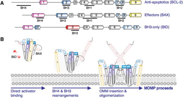
(A) The BCL-2 proteins are comprised of BCL-2 homology (BH) domains. A representation of an antiapoptotic (BCL-2), effector (BAX), and BH3-only (BID) protein is shown with the BH1-4 designated underneath the corresponding α helices. (B) Proposed model of BAX activation. Soluble BAX interacts with a direct activator and the OMM to promote stable N-terminal exposure, and BAX α5, α6, and α9 insert within the OMM.
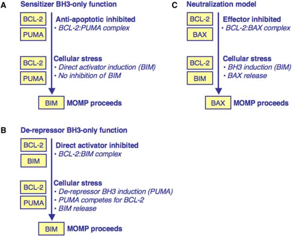
(A) Sensitizer BH3-only protein function. A sensitizer BH3-only protein inhibits the antiapoptotic BCL-2 repertoire. Following minimal cellular stress, a direct activator is induced but cannot be inhibited and MOMP proceeds. (B) Derepressor BH3-only protein function. A direct activator is sequestered by an antiapoptotic BCL-2 protein. Following cellular stress, a derepressor BH3-only protein is induced and competes with the direct activator for binding to the antiapoptotic repertoire. When the direct activator is released, MOMP proceeds. (C) The neutralization model of BCL-2 family function. In this model, BAK/BAX are always competent to promote MOMP but are actively inhibited by the antiapoptotic BCL-2 repertoire to promote survival. Following cellular stress, BH3-only proteins are induced, bind the antiapoptotic proteins, and displace effectors to promote MOMP.
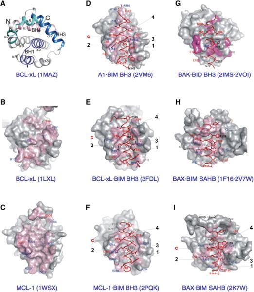
(A) The “front” view of BCL-xL identifies the BCL-2 core and the respective locations of the four BH regions. The PDB identifier is in parentheses. (B) Surface representation of free BCL-xL, emphasizing amino acids participating in BH3 peptide binding at the “front” face, identifies the BC groove. Representations are partially transparent, permitting identification, beneath the surface, of contact side chains positioned 4Å from a peptide. With the exception of free BCL-xL and MCL-1 (C), both having surface coloring based on all atoms of peptide amino acids, surface coloring highlights strictly the atoms within 4Å from a peptide. PDB residue numbering has been maintained. Labels of acidic and basic amino acids are colored red and blue, respectively. (C) Free MCL-1. (D) A1 bound to BIM BH3. The four conserved hydrophobic residues/sites and the conserved charged interactions are marked. Peptide amino acid labels are bold. (E) BCL-xL bound to BIM BH3. (F) MCL-1 bound to BIM BH3. (G) A model between BID BH3 (from the A1·BID BH3 complex) and BAK-ΔTM, overlapping sites of interaction (colored) identified by NMR spectroscopy. (H) BIM SAHB modeling to the “back” face of free BAX. (I) BIM SAHB binding site on BAX.
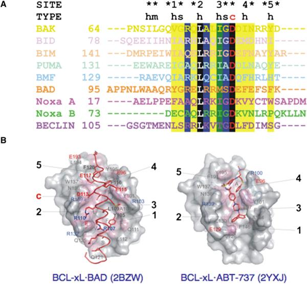
(A) Structure-based alignment of BH3 peptides from complexes with A1 (BAK, 2VOH; BID, 2VOI; BIM, 2VM6; PUMA, 2VOH; BMF, 2VOG), BCL-xL (2BZW, full-length BAD; 2P1L, BECLIN), and MCL-1 (Noxa A, 2ROD; Noxa B, 2NLA; see Table S2). BC grooves accommodate different types of BH3 amino acids: h, hydrophobic; m, mixed; s, small; c, charged. The degree of conservation, illustrated in black (100%), green (>75%), blue (>50%), and yellow (25%), identifies a conserved core defining BH3 peptide specificity. The five conserved hydrophobic residues/sites and the conserved charged interactions are marked. (B) Structure of BCL-xL illustrates overlapping sites of interaction with full-length BAD and ABT-737.
References
-
- Arnoult D, Grodet A, Lee YJ, Estaquier J, Blackstone C. Release of OPA1 during apoptosis participates in the rapid and complete release of cytochrome c and subsequent mitochondrial fragmentation. J. Biol. Chem. 2005;280:35742–35750. - PubMed
-
- Autret A, Martin SJ. Emerging role for members of the Bcl-2 family in mitochondrial morphogenesis. Mol. Cell. 2009;36:355–363. - PubMed
-
- Bedikian AY, Millward M, Pehamberger H, Conry R, Gore M, Trefzer U, Pavlick AC, DeConti R, Hersh EM, Hersey P, et al. Bcl-2 antisense (oblimersen sodium) plus dacarbazine in patients with advanced melanoma: the Oblimersen Melanoma Study Group. J. Clin. Oncol. 2006;24:4738–4745. - PubMed
-
- Cassidy-Stone A, Chipuk JE, Ingerman E, Song C, Yoo C, Kuwana T, Kurth MJ, Shaw JT, Hinshaw JE, Green DR, Nunnari J. Chemical inhibition of the mitochondrial division dynamin reveals its role in Bax/Bak-dependent mitochondrial outer membrane permeabilization. Dev. Cell. 2008;14:193–204. - PMC - PubMed
Publication types
MeSH terms
Substances
Grants and funding
LinkOut - more resources
Full Text Sources
Other Literature Sources
