. 2010 Feb 16;18(2):214-25.
doi: 10.1016/j.devcel.2010.01.007.

Structural basis of selective ubiquitination of TRF1 by SCFFbx4

Affiliations

Structural basis of selective ubiquitination of TRF1 by SCFFbx4

Zhixiong Zeng et al. Dev Cell. .

Abstract

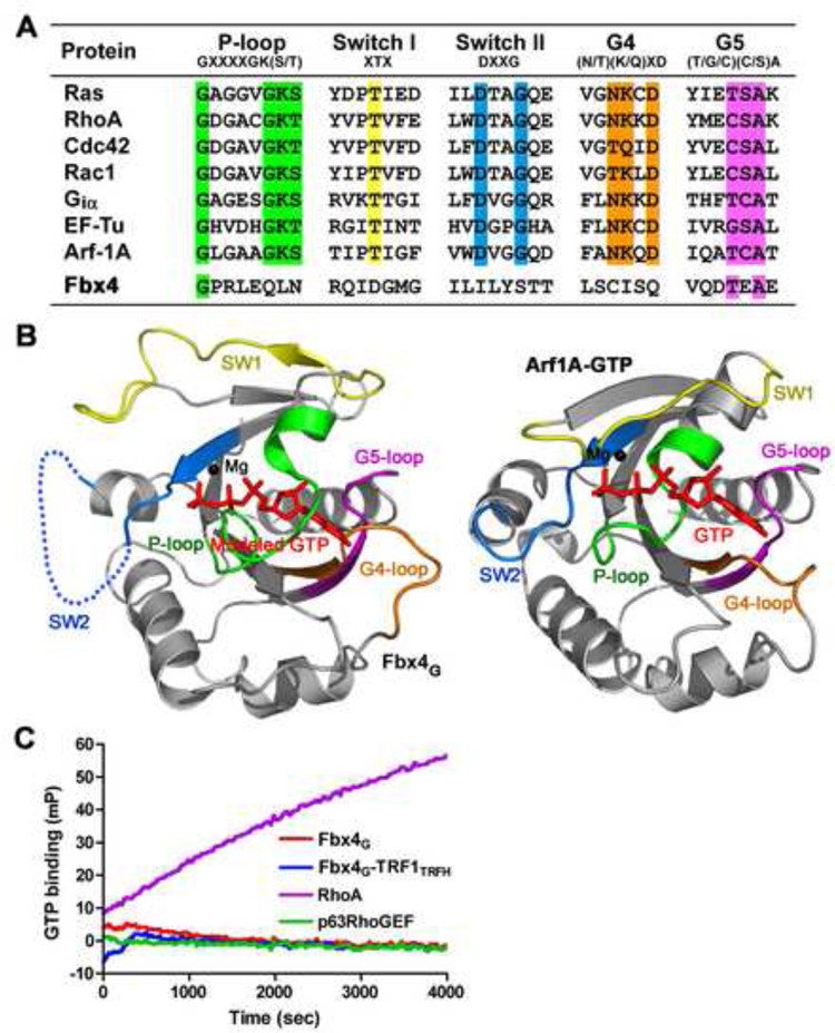
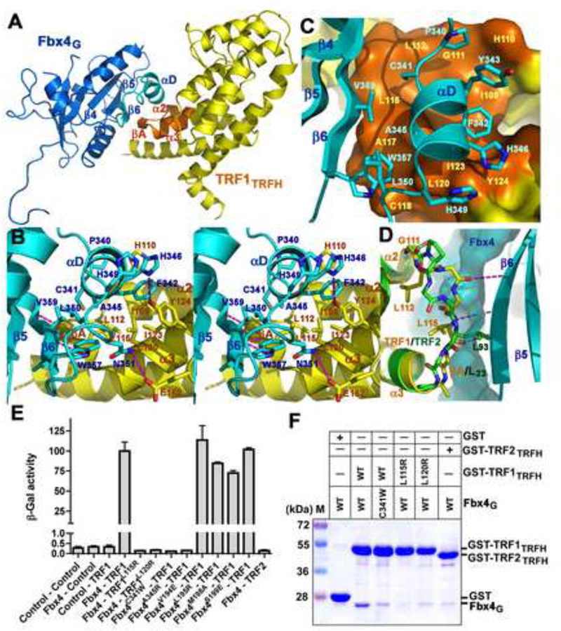
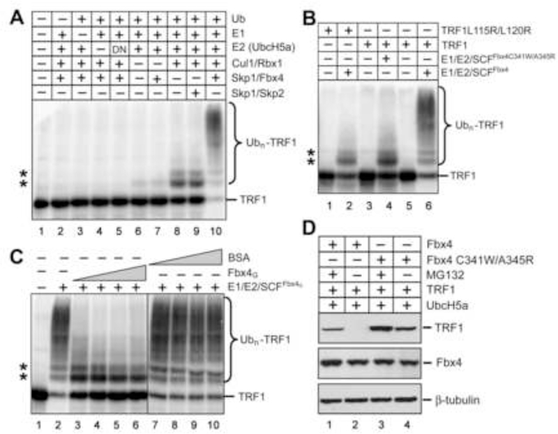
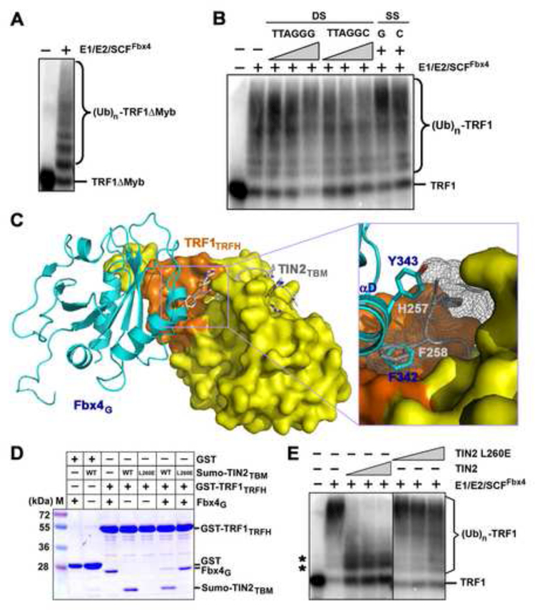
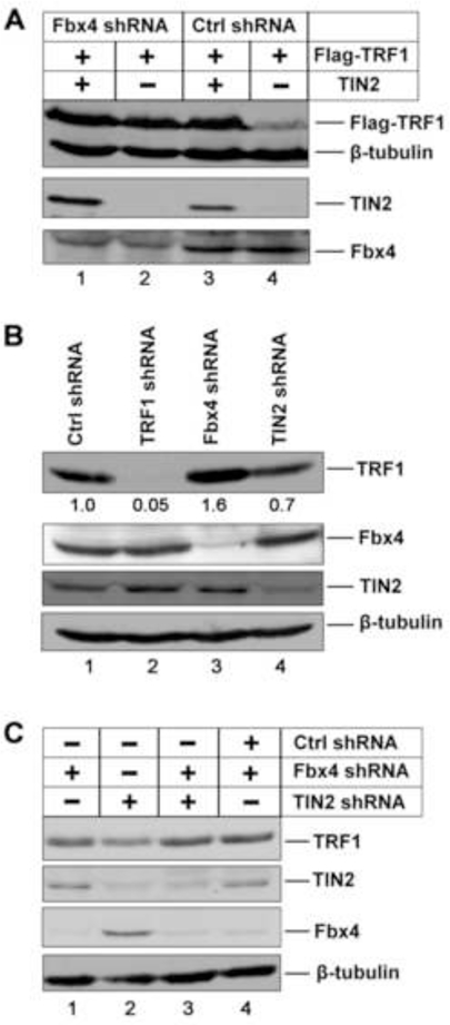
TRF1 is a critical regulator of telomere length. As such, TRF1 levels are regulated by ubiquitin-dependent proteolysis via an SCF E3 ligase where Fbx4 contributes to substrate specification. Here, we report the crystal structure of the Fbx4-TRF1 complex at 2.4 A resolution. Fbx4 contains an unusual substrate-binding domain that adopts a small GTPase fold. Strikingly, this atypical GTPase domain of Fbx4 binds to a globular domain of TRF1 through an intermolecular beta sheet, instead of recognizing short peptides/degrons as often seen in other F-box protein-substrate complexes. Importantly, mutations in this interface abrogate Fbx4-dependent TRF1 binding and ubiquitination. Furthermore, the data demonstrate that recognition of TRF1 by SCF(Fbx4) is regulated by another telomere protein, TIN2. Our results reveal an atypical small GTPase domain within Fbx4 as a substrate-binding motif for SCF(Fbx4) and uncover a mechanism for selective ubiquitination and degradation of TRF1 in telomere homeostasis control.


Figures

Figure 1.
Figure 1.. Overview of the Fbx4G-TRF1TRFH Complex Structure
(A) Domain organization of the Fbx4 and TRF1 polypeptide chains. In Fbx4, the F-box is colored in green and the C-terminal Fbx4G domain in cyan. In TRF1, the N-terminal acidic region is in red, the C-terminal Myb domain in slate, and the TRFH domain in yellow. The shaded area between Fbx4 and TRF1 indicates that the Fbx4-TRF1 interaction is mediated by Fbx4G and TRF1TRFH. (B) Ribbon diagram of the dimeric Fbx4G-TRF1TRFH complex. Fbx4G and TRF1TRFH are colored in cyan and yellow, respectively, in one complex, and blue and orange in the other. The secondary structure elements are labeled. (C) Surface representation indicates that the dimeric Fbx4G-TRF1TRFH complex adopts a saddle-shaped conformation. The orientation of complex is rotated by 90° about a horizontal axis relative to the complex in (B).
Figure 2.
Figure 2.. Fbx4G is an Atypical Small GTPase Domain without GTP Binding Activity
(A) Sequence alignment in the loop regions of Fbx4G and a group of small GTPases. The conserved residues important for nucleotide binding are highlighted in different colors;the P-loop is in green, Switch I in yellow, Switch II in cyan, G4 in orange and G5 in magenta. (B) The conformation of Fbx4G observed in the crystal structure is incompatible with GTP binding. Left: ribbon representation of Fbx4G with modeled GTP and Mg2+. The Ploop, Switch II and the G4-loop of Fbx4G block the binding of GTP. Right: ribbon representation of the Arf1A-GTP complex (PDB: 2J59). The coloring scheme of the loop regions is the same as in (A). GTP is shown as a stick model and Mg2+ a black ball. (C) Times courses of GTP binding for Fbx4G and the Fbx4G-TRF1TRFH complex. GTP binding was monitored by the increase in fluorescence millipolarization (mP) of a fluorescent GTP analog as it was bound. In this experiment, neither Fbx4G nor the Fbx4GTRF1TRFH complex bound to GTP. RhoA (a small GTPase) and p63RhoGEF (a non GTPase protein) were used as positive and negative controls, respectively.
Figure 3.
Figure 3.. Recognition of TRF1 by Fbx4
(A) An overall view of the Fbx4G-TRF1TRFH interaction. The interacting structural elements of Fbx4G (β6 and αD) and TRF1TRFH (βA, α2 and α3) are colored in cyan and orange, respectively, and the rest of Fbx4G and TRF1TRFH in blue and yellow. (B) Stereo view of the Fbx4G-TRF1TRFH interface. Fbx4G and TRF1TRFH interacting residues are presented as stick models. The Fbx4G-TRF1TRFH intermolecular hydrogen bonds are shown as dashed magenta lines. (C) The αD helix of Fbx4G sits on a hydrophobic surface formed by μA and helices of α2 and α3 of TRF1TRFH. (D) Fbx4G binding is TRF1TRFH specific. Loop L23 of TRF2TRFH (green) adopts a different conformation than that of TRF1TRFH (yellow) so that it cannot form an intermolecular β-sheet interaction with Fbx4G. The surface representation shows that the side chain of TRF2 Leu93 collides with μ6 of Fbx4G. (E) Effects of the Fbx4 and TRF1 mutations on the Fbx4-TRF1 interaction in a yeast two-hybrid assay. Interaction of LexA-TRF1 with GAD-Fbx4 was measured as β-galactosidase activity. Data are average of three independent β-galactosidase measurements normalized to the wild-type Fbx4-TRF1 interaction, arbitrarily set to 100. Error bars in the graph represent standard deviation. (F) GST pull-down assay of the wild-type and mutant Fbx4G-TRF1TRFH interactions.
Figure 4.
Figure 4.. The Fbx4-TRF1 Interface Is Essential for Fbx4-dependent TRF1 Ubiquitination
(A) Fbx4-dependent of ubiquitination of TRF1. Purified recombinant TRF1 was 33Plabeled by protein kinase GST-cyclin B/Cdk1 and 33P-TRF1 was separated from GSTcyclin B/Cdk1 by depletion with GSH beads. 33P-TRF1 was then incubated with purified recombinant E1, E2(UbcH5a), E3(SCFFbx4) enzymes and ubiquitin, methylated ubiquitin, ubiquitin aldehyde and MG 132. Reaction mixtures were separated by SDS-PAGE, followed by PhosphorImaging analysis. In panels (A), (B) and (C), asterisks indicate the non-specific ubiquitination products of TRF1 due to the presence of Cul1/Rbx1. (B) The Fbx4-TRF1 interface is essential for Fbx4-dependent ubiquitination of TRF1. Reactions were performed as in (A), but TRF1L115R/L120R was used as substrate in one reaction and SCFFbx4C341W/A345R as E3 ligase in another. (C) Addition of Fbx4G suppresses Fbx4-dependent ubiquitination of TRF1. Ubiquitination assays were performed as in (A), but increasing amount of Fbx4G (3, 6, 12, 24 μM) or BSA (3, 6, 12, 24 μM) was added in the reactions. (D) HEK 293T cells were transfected with TRF1, UbcH5a, Fbx4 WT or Fbx4 C341W/A345R. Four hours after transfection, cells were split into two plates and grown for 24 hours prior to being treated with 2 µM MG 132 or DMSO for 16 hours. Cells were lysed and cell extracts were immunoblotted with indicated antibodies.
Figure 5.
Figure 5.. TIN2 Blocks the Fbx4-TRF1 Interaction and Inhibits the Ubiquitination of TRF1 Mediated by SCFFbx4
(A) TRF1 deletion mutant lacking the DNA-binding Myb domain, TRF1ΔMyb, can be efficiently ubiquitinated by SCFFbx4. (B) Addition of either wt or mutant telomere DNAs has no effect on TRF1 ubiquitination. In vitro ubiquitination reactions containing 33P-labeled TRF1 were carried out in the absence (−) or presence of double-stranded (DS; 1, 12 or 36 μM TTAGGG or TTAGGC) repeat DNA or single-stranded [SS; 36 μM TTAGGG (G) or AATCCC (C)] repeat DNA. (C) Superposition of the Fbx4G-TRF1TRFH structure with the crystal structure of the TRF1TRFH-TIN2TBM complex. Fbx4G (cyan) and TIN2TBM (gray) share a common hydrophobic interacting surface on TRF1TRFH (orange). An enlarged view of the collision between Fbx4G αD helix and the TIN2TBM peptide is highlighted in the box. (D) GST pull-down competition assay of the Fbx4G-TRF1TRFH interaction in the presence of wt or the L260E mutant TIN2TBM peptides. For clarity, Sumo-fused TIN2TBM peptides were used in this assay. Sumo itself does not bind to TRF1TRFH (data not shown). (E) TIN2 inhibits Fbx4 mediated ubiquitination of TRF1. TRF1 ubiquitination assays were performed as in (A), but increasing amount of TIN2 (3, 9, 27, 81 μM) or mutant TIN2L260E (3, 9, 27, 81 μM) was added in the reactions. Asterisks indicate the non-specific ubiquitination products of TRF1 due to the presence of Cul1/Rbx1.
Figure 6.
Figure 6.. TRF1 stabilization by TIN2 depends on Fbx4
(A) HeLa cells expressing control or Fbx4 shRNA were transfected with Flag-TRF1 along with either TIN2 or an empty expression vector. The expression levels of Flag-TRF1, TIN2 and Fbx4 were analyzed Immunoblotting with the respective antibodies. β-tubulin was blotted as the loading control. (B) Immunoblotting analysis of the endogenous levels of TRF1 in HeLa cells stably expressing control, TRF1, Fbx4 or TIN2 shRNA vectors with the indicated antibodies. (C) Endogenous levels of TRF1 in Fbx4 or TIN2 single or double knockdown cells as determined by immunoblotting with the indicated antibodies.

Comment in

  • Telomeric TuRF1 wars.
    Sarraf SA, Harper JW. Sarraf SA, et al. Dev Cell. 2010 Feb 16;18(2):167-8. doi: 10.1016/j.devcel.2010.02.002. Dev Cell. 2010. PMID: 20159586

Similar articles

Cited by

References

    1. Aspenstrom P, Ruusala A, and Pacholsky D (2007). Taking Rho GTPases to the next level: the cellular functions of atypical Rho GTPases. Experimental Cell Research 313, 3673–3679. - PubMed
    1. Atanassov BS, Evrard YA, Multani AS, Zhang Z, Tora L, Devys D, Chang S, and Dent SY (2009). Gcn5 and SAGA regulate shelterin protein turnover and telomere maintenance. Molecular Cell 35, 352–364. - PMC - PubMed
    1. Blackburn EH (2001). Switching and signaling at the telomere. Cell 106, 661–673. - PubMed
    1. Broccoli D, Smogorzewska A, Chong L, and de Lange T (1997). Human telomeres contain two distinct Myb-related proteins, TRF1 and TRF2. Nature Genetics 17, 231–235. - PubMed
    1. Brunger AT, Adams PD, Clore GM, DeLano WL, Gros P, Grosse-Kunstleve RW, Jiang JS, Kuszewski J, Nilges M, Pannu NS, et al. (1998). Crystallography & NMR system: A new software suite for macromolecular structure determination. Acta Crystallogr D Biol Crystallogr 54 (Pt 5), 905–921. - PubMed

Publication types

MeSH terms

Substances

Associated data