. 2010 Feb 16;18(2):226-36.
doi: 10.1016/j.devcel.2009.11.015.

Differential regulation of protrusion and polarity by PI3K during neutrophil motility in live zebrafish

Affiliations

Differential regulation of protrusion and polarity by PI3K during neutrophil motility in live zebrafish

Sa Kan Yoo et al. Dev Cell. .

Erratum in

  • Dev Cell. 2011 Aug 16;21(2):384

Abstract

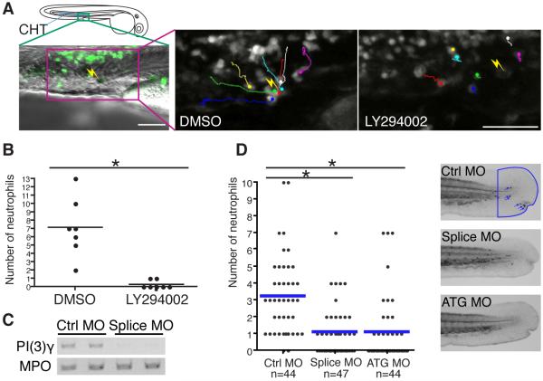
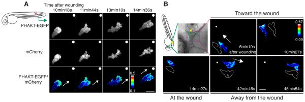
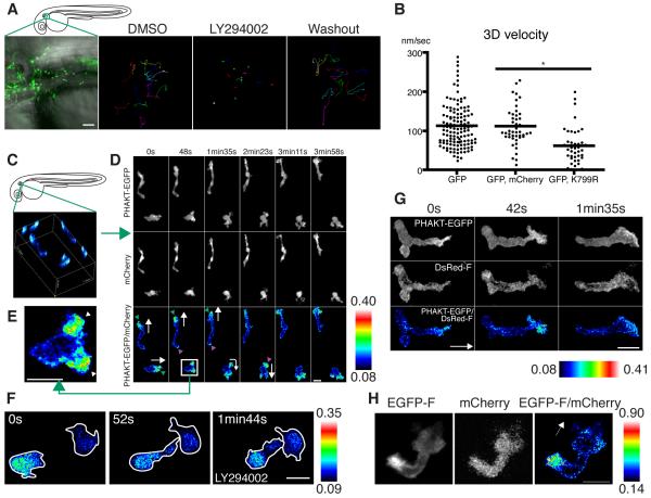
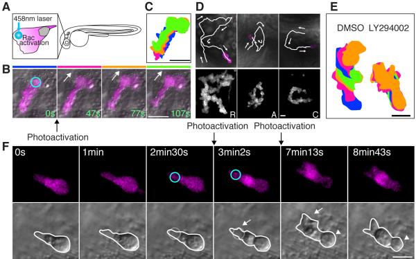
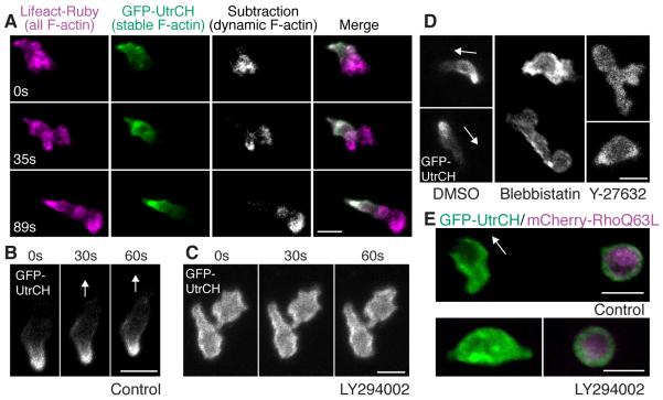
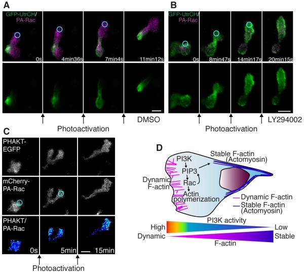
Cell polarity is crucial for directed migration. Here we show that phosphoinositide 3-kinase (PI(3)K) mediates neutrophil migration in vivo by differentially regulating cell protrusion and polarity. The dynamics of PI(3)K products PI(3,4,5)P(3)-PI(3,4)P(2) during neutrophil migration were visualized in living zebrafish, revealing that PI(3)K activation at the leading edge is critical for neutrophil motility in intact tissues. A genetically encoded photoactivatable Rac was used to demonstrate that localized activation of Rac is sufficient to direct migration with precise temporal and spatial control in vivo. Similar stimulation of PI(3)K-inhibited cells did not direct migration. Localized Rac activation rescued membrane protrusion but not anteroposterior polarization of F-actin dynamics of PI(3)K-inhibited cells. Uncoupling Rac-mediated protrusion and polarization suggests a paradigm of two-tiered PI(3)K-mediated regulation of cell motility. This work provides new insight into how cell signaling at the front and back of the cell is coordinated during polarized cell migration in intact tissues within a multicellular organism.


Figures

Figure 1
Figure 1
PI(3)Kγ is necessary for directed migration of neutrophils in vivo. (A) 65 μM LY294002 treatment inhibits attraction of neutrophils to a laser wound in the caudal hematopoietic tissue (CHT). The lines indicate tracking of individual neutrophils over 30 minutes and the yellow thunder shows the position of the laser wound. Note neutrophils migrate rapidly toward the wound in control (DMSO), but not after LY294002 treatment (movie S1). (B) The number of neutrophils that reach the laser wound in the CHT within 30 minutes is quantified (7 movies for each condition, *, P<0.001, two-tailed unpaired t-test). Note LY294002 treatment inhibits attraction of neutrophils to the wound. (C) Splice morpholino disturbs splicing of PI(3)Kγ transcript. MPO transcript indicates integrity of neutrophils in morphants. (D) Knockdown of PI(3)Kγ inhibits directional migration of neutrophils to wounds (neutrophils are indicated with blue arrows in control, *, P<0.001, two-tailed unpaired t-test). Data of (D) are representative of 3 separate experiments. Scale bar, 50 μm (A).
Figure 2
Figure 2
PHAKT-EGFP translocates to the leading edge when neutrophils come to and leave laser-induced wounds. (A) Time-lapse ratiometric imaging (PHAKT-EGFP/mCherry) reveals PI(3,4,5)P3-PI(3,4)P2 localization at the leading edge during attraction to a laser wound in the tail fin (movie S2A). The white dots and arrows indicate the position of the wound and direction of migration respectively. (B) Reversal of PI(3,4,5)P3-PI(3,4)P2 (ratiometric imaging of PHAKT-EGFP/mCherry) when a neutrophil leaves the laser wound (movie S2B). Note loss of PI(3,4,5)P3-PI(3,4)P2 polarity at the wound, followed by reversal of polarity to the opposite pole away from the wound when the neutrophil leaves the wound (green line: tracking of a neutrophil, yellow thunder: position of the laser wound, white arrows: direction of migration, illustration with white line: morphology of a neutrophil). Data are representative of more than 5 time-lapse movies from a minimum of 3 separate experiments. The numerical values of ratiometric analysis are shown in the scales. Scale bars, 10 μm (A, B).
Figure 3
Figure 3
PI(3)K is critical for neutrophil motility and is active at the leading edge in the mesenchymal tissues of the head. (A) Random migration of neutrophils is arrested by 65 μM LY294002 and restored after washout of the drug. The lines indicate tracking of neutrophil motility (12 cells per condition) imaged for 30 minutes using Tg(MPO:GFP)uw (movie S3A). (B) PI(3)Kγ K799R disturbs interstitial motility of neutrophils (movie S3B, *, P<0.001, two-tailed unpaired t-test, GFP: 128 neutrophils (40 movies), GFP, mCherry: 45 neutrophils (9 movies), GFP, K799R: 44 neutrophils (34 movies). (C) 3D reconstruction of ratiometric image (PHAKT-EGFP/mCherry). (D) Time-lapse ratiometric imaging (PHAKT-EGFP/mCherry) of PI(3,4,5)P3-PI(3,4)P2 dynamics during random migration (movie S4A). PI(3,4,5)P3-PI(3,4)P2 is mainly localized at the leading edge (green arrowheads) and occasionally at the tail (magenta arrow heads). White arrows indicate direction of migration. (E) PI(3,4,5)P3-PI(3,4)P2 at the bifurcated pseudopod, indicated by arrowheads (movie S4A). (F) Treatment with 65 μM LY294002 inhibits the leading edge signal of PI(3,4,5)P3-PI(3,4)P2 and induces high ratiometric signals of PHAKT-EGFP/mCherry in the cell body of neutrophils (movie S4C). Note the rounded tails and thin pseudopods induced by LY294002. (G) PI(3,4,5)P3-PI(3,4)P2 signal at the leading edge by ratiometric imaging of PHAKT-EGFP/farnesylated DsRed (DsRed-F) (movie S4D). The white arrow indicates direction of migration. (H) Ratiometric imaging of EGFP-F/mCherry reveals periodic accumulation of membrane components at the tail (movie S4E). The white arrow indicates direction of migration. Images are representative of 3 (A) and more than 5 (C-H) time-lapse movies from a minimum of 3 separate experiments. Scale bars, 50 μm (A), 10 μm (D-H).
Figure 4
Figure 4
Photoactivation of Rac at the leading edge can rescue the protrusion defects but not the rounded tail or migration defects induced by PI(3)K inhibition. (A) A schematic representation of photoactivation of Rac at the neutrophil leading edge in zebrafish. (B) Photoactivation of Rac at the leading edge induces protrusion and migration of a neutrophil in tissues (movie S5). The circle indicates the position of Rac photoactivation. (C) Overlayed images of (B) show directional migration induced by photoactivated Rac. (D) Spelling by neutrophil trajectories guided through repetitive photoactivation of Rac at the leading edge (movie S6). (E) Overlayed images show that PI(3)K inhibition disturbs Rac photoactivation-induced migration (The leading edge was activated for 20 seconds twice during 5 minute imaging). (F) Photoactivation of Rac at the front (circles) can rescue the protrusion defect induced by PI(3)K inhibition (arrows), but not the rounded tail defect (arrowheads) (movie S8). Images are representative of more than 5 time-lapse movies from experiments repeated on at least two separate dates. Scale bars, 20 μm (B, C), 10 μm (D-F).
Figure 5
Figure 5
PI(3)K regulates anteroposterior polarity of F-actin dynamics. (A) Stable F-actin (GFP-UtrCH) is localized at the tail while dynamic F-actin (Lifeact-Ruby subtracted by GFP-UtrCH) is localized at the front (movie S9). (B) In control, GFP-UtrCH labels the tail. (C) PI(3)K inhibition by LY294002 disturbs tail localization of stable F-actin (movie S11A). (D) Myosin ATPase and Rho kinase inhibition disturbs tail localization of stable F-actin (movie S11C). (E) Constitutively active RhoQ63L induces cell rounding and localization of stable F-actin all over the membrane (top panel). PI(3)K inhibition does not relieve constitutively active Rho-mediated effects on cell rounding or localization of stable F-actin (lower panel) (movie S12A). Images are representative of more than 5 time-lapse movies from experiments repeated on at least two separate dates. Scale bars, 10 μm.
Figure 6
Figure 6
PI(3)K regulates anteroposterior polarity of F-actin dynamics in a pathway that is separable from Rac-mediated protrusion. (A) In control, photoactivation of Rac at the leading edge induces protrusion at the leading edge with GFP-UtrCH (stable F-actin) localized at the tail (movie S13A). (B) Photoactivation of Rac with PI(3)K inhibition induces protrusion with GFP-UtrCH (stable F-actin) localized at the leading edge, indicating reverse polarity of F-actin dynamics (movie S13B). (C) Protrusion induced by photoactivation of Rac induces PHAKT-EGFP accumulation at the leading edge, suggesting a positive feedback from Rac to PI(3,4,5)P3-PI(3,4)P2 gradient (movie S14). (D) A schematic representation of two-tiered PI(3)K-mediated regulation of cell motility: PI(3)K promotes Rac-mediated actin polymerization at the leading edge while generating anteroposterior polarity of F-actin dynamics. Images are representative of more than 5 time-lapse movies from experiments repeated on at least two separate dates. Scale bars, 10 μm.

Comment in

Similar articles

Cited by

References

    1. Andrew N, Insall RH. Chemotaxis in shallow gradients is mediated independently of PtdIns 3-kinase by biased choices between random protrusions. Nat Cell Biol. 2007;9:193–200. - PubMed
    1. Arrieumerlou C, Meyer T. A local coupling model and compass parameter for eukaryotic chemotaxis. Dev Cell. 2005;8:215–227. - PubMed
    1. Barberis L, Hirsch E. Targeting phosphoinositide 3-kinase gamma to fight inflammation and more. Thromb Haemost. 2008;99:279–285. - PubMed
    1. Benink HA, Bement WM. Concentric zones of active RhoA and Cdc42 around single cell wounds. J Cell Biol. 2005;168:429–439. - PMC - PubMed
    1. Burkel BM, von Dassow G, Bement WM. Versatile fluorescent probes for actin filaments based on the actin-binding domain of utrophin. Cell Motil Cytoskeleton. 2007;64:822–832. - PMC - PubMed

Publication types

MeSH terms

Substances