

Free-breathing diffusion-weighted single-shot echo-planar MR imaging using parallel imaging (GRAPPA 2) and high b value for the detection of primary rectal adenocarcinoma
- PMID: 20159662
- PMCID: PMC2842173
- DOI: 10.1102/1470-7330.2010.0011
Free-breathing diffusion-weighted single-shot echo-planar MR imaging using parallel imaging (GRAPPA 2) and high b value for the detection of primary rectal adenocarcinoma
Abstract
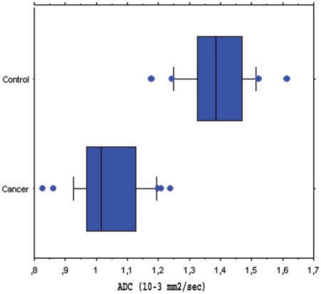
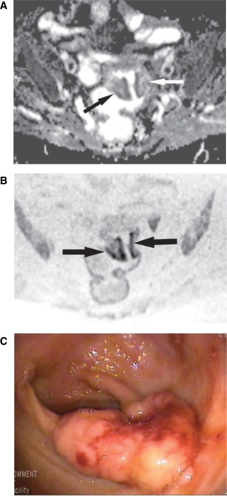
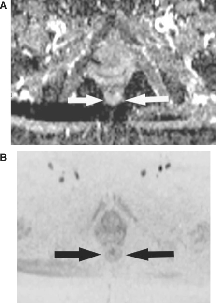
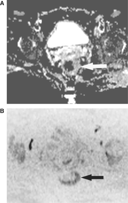
Our objective was to determine the diagnostic accuracy of a free-breathing diffusion-weighted single-shot echo-planar magnetic resonance imaging (FBDW-SSEPI) technique with parallel imaging and high diffusion factor value (b = 1000 s/mm2) in the detection of primary rectal adenocarcinomas. Thirty-one patients (14M and 17F; mean age 67 years) with histopathologically proven primary rectal adenocarcinomas and 31 patients without rectal malignancies (14M and 17F; mean age 63.6 years) were examined with FBDW-SSEPI (repetition time (TR/echo time (TE) 3900/91 ms, gradient strength 45 mT/m, acquisition time 2 min) at 1.5 T using generalized autocalibrating partially parallel acquisitions (GRAPPA, acceleration factor 2) and a b value of 1000 s/mm2. Apparent diffusion coefficients (ADCs) of rectal adenocarcinomas and normal rectal wall were measured. FBDW-SSEPI images were evaluated for tumour detection by 2 readers. Sensitivity, specificity, accuracy and Youden score for rectal adenocarcinoma detection were calculated with their 95% confidence intervals (CI) for ADC value measurement and visual image analysis. Rectal adenocarcinomas had significantly lower ADCs (mean 1.036 x 10(-3)+/- 0.107 x 10(-3) mm2/s; median 1.015 x 10(-3) mm2/s; range (0.827-1.239) x 10(-3) mm2/s) compared with the rectal wall of control subjects (mean 1.387 x 10(-3)+/- 0.106 x 10(-3) mm2/s; median 1.385 x 10(-3) mm2/s; range (1.176-1.612) x 10(-3) mm2/s) (p < 0.0001). Using a threshold value < or = 1.240 x 10(-3) mm2/s, all rectal adenocarcinomas were correctly categorized and 100% sensitivity (31/31; 95% CI 95-100%), 94% specificity (31/33; 95% CI 88-100%), 97% accuracy (60/62; 95% CI 92-100%) and Youden index 0.94 were obtained for the diagnosis of rectal adenocarcinoma. FBDW-SSEPI image analysis allowed depiction of all rectal adenocarcinomas but resulted in 2 false-positive findings, yielding 100% sensitivity (31/31; 95% CI 95-100%), 94% specificity (31/33; 95% CI 88-100%), 97% accuracy (60/62; 95% CI 92-100%) and Youden index 0.94 for the diagnosis of primary rectal adenocarcinoma. We can conclude that FBDW-SSEPI using parallel imaging and high b value may be helpful in the detection of primary rectal adenocarcinomas.
Figures
References
-
- Ichikawa T, Haradome H, Hachiya J, Nitatori T, Araki T. Diffusion-weighted MR imaging with single-shot echo-planar imaging in the upper abdomen: preliminary clinical experience in 61 patients. Abdom Imaging. 1999;24:456–61. doi:10.1007/s002619900539. PMid:10475927. - DOI - PubMed
-
- Ichikawa T, Ertuk SM, Motosugi U, et al. High-b-value diffusion-weighted MRI in colorectal cancer. AJR Am J Roentgenol. 2006;187:181–4. doi:10.2214/AJR.05.1005. PMid:16794174. - DOI - PubMed
-
- Ichikawa T, Haradome H, Hachiya J, Nitatori T, Araki T. Diffusion-weighted MR imaging with a single-shot echoplanar sequence: detection and characterization of focal hepatic lesions. AJR Am J Roentgenol. 1998;170:397–402. - PubMed
-
- Taouli B, Martin AJ, Qayyum A, et al. Parallel imaging and diffusion tensor imaging for diffusion-weighted MRI of the liver: preliminary experience in healthy volunteers. AJR Am J Roentgenol. 2004;183:677–80. - PubMed
-
- Rao SX, Zeng MS, Chen CZ, et al. The value of diffusion-weighted imaging in combination with T2-weighted imaging for rectal cancer detection. Eur J Radiol. 2008;65:299–303. doi:10.1016/j.ejrad.2007.04.001. PMid:17498902. - DOI - PubMed
MeSH terms
LinkOut - more resources
Full Text Sources