. 2010 May;77(5):874-83.
doi: 10.1124/mol.109.060301. Epub 2010 Feb 16.

Increased insertion of glutamate receptor 2-lacking alpha-amino-3-hydroxy-5-methyl-4-isoxazole propionic acid (AMPA) receptors at hippocampal synapses upon repeated morphine administration

Affiliations

Increased insertion of glutamate receptor 2-lacking alpha-amino-3-hydroxy-5-methyl-4-isoxazole propionic acid (AMPA) receptors at hippocampal synapses upon repeated morphine administration

Sophie K Billa et al. Mol Pharmacol. 2010 May.

Abstract

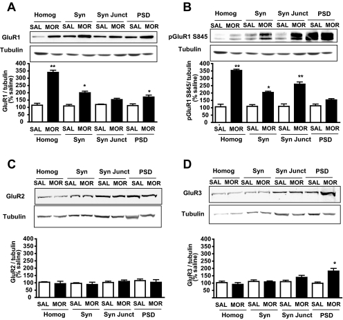
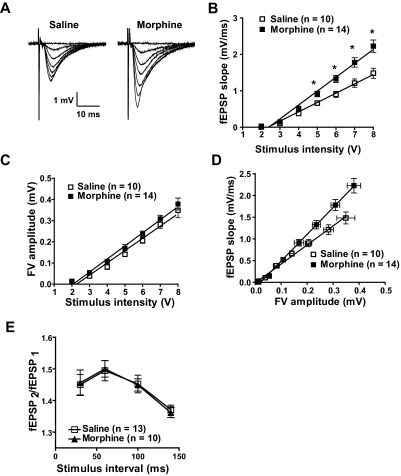
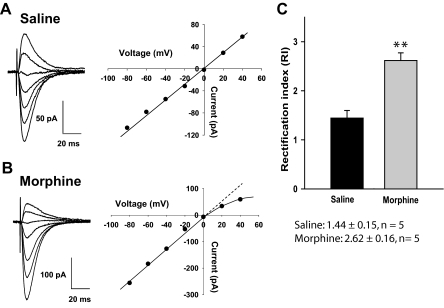
Evidence suggests that the long-term adaptations in the hippocampus after repeated drug treatment may parallel its role during memory formation. The neuroplasticity that subserves learning and memory is also believed to underlie addictive processes. We have reported previously that repeated morphine administration alters local distribution of endocytic proteins at hippocampal synapses, which could in turn affect expression of glutamate receptors. Glutamatergic systems, including alpha-amino-3-hydroxy-5-methyl-4-isoxazole propionic acid receptors (AMPARs), are believed to be involved in opiate-induced neuronal and behavioral plasticity, although the mechanisms underlying these effects are only beginning to be understood. The present study further examines the effects of repeated morphine administration on the expression and composition of AMPARs and the functional ramifications. Twelve hours after the last morphine injection, we observed an increased expression of AMPARs lacking glutamate receptor (GluR) 2 in hippocampal synaptic fractions. Immunoblotting studies show that 12 h after morphine treatment, GluR1 subunits are increased at the postsynaptic density (PSD) and at extrasynaptic sites, whereas GluR3 subunits are only increased at the PSD, and they show how this alters receptor subunit composition. In addition, we provide electrophysiological evidence that AMPARs are switched to Ca(2+)-permeable (GluR2-lacking) at the synapse 12 h after repeated morphine treatment, affecting the magnitude of long-term depression at hippocampal neurons. We propose that morphine-induced changes in glutamatergic synaptic transmission in the hippocampus may play an important role in the neuroadaptations induced by repeated morphine administration.


Figures

Fig. 1.
Fig. 1.
Morphine-induced expression changes of AMPA subunits in hippocampal synaptic fractions 12 h after treatment. Fractions representing homogenate (Homog), synaptosomes (Syn), synaptic junctions (Syn Jct), and PSD were subjected to Western blot analyses. Morphine treatment increased levels of GluR1 at the homogenate, the synaptosomal fraction, and the PSD (A). Phosphorylation of GluR1 at Ser845 was also increased at the homogenate, synaptosomes, and synaptic junctions (B). In contrast, GluR2 levels were not altered upon morphine treatment (C). Morphine treatment induced an increase of GluR3 levels at the PSD (D). Blots were standardized with tubulin. Quantification was performed relative to tubulin levels (*, p < 0.05, **, p < 0.01 relative to saline-treated animals, one-way ANOVA with Tukey's multiple comparison test, n = 6). SAL, saline; MOR, morphine.
Fig. 2.
Fig. 2.
Quantitative coimmunoprecipitation of AMPA subunits in hippocampal synaptosomal membranes 12 h after repeated morphine administration. AMPAR subunit composition was compared in hippocampal synaptosomal membranes from saline- and morphine-treated mice 12 h after discontinuation of morphine treatment. Immunoblots (IB) show the percentage of AMPAR subunits remaining (unbound fraction) after IP of synaptosomal membranes. Data shown represent the average of two independent experiments. The antibodies used for IB are indicated at the left. The left three lanes in each row show immunoblotting of IgG control immunoprecipitated sample. After IP with the respective antibody, the percentage remaining in the unbound fraction was calculated from the standard curve generated by controls of 100 (14 μg), 50 (7 μg), and 5% (0.7 μg) run on each blot. Results obtained indicate that morphine administration decreases the association between GluR1 and GluR2 and GluR3 and increases the association between GluR2 and GluR3.
Fig. 3.
Fig. 3.
Morphine increases fEPSP but not fiber volley magnitude. A, traces of FVs and fEPSPs were recorded 12 h after discontinuation of treatment in hippocampal slices from saline- and morphine-treated animals. B, fEPSP magnitude increased as a function of stimulus intensity and was greater in morphine-treated animals (F1,126 = 9.25, *, p < 0.05, two-way ANOVA). C, relationship between FV amplitude and stimulus intensity did not differ between saline- and morphine-treated animals. D, input (FV)-output (fEPSP) shows that morphine treatment increased fEPSP magnitude at similar presynaptic fiber volley amplitudes (linear regression comparison of fit of two curves, F3,294 = 31.8, p < 0.0001). E, PPF does not differ between saline- and morphine-treated animals.
Fig. 4.
Fig. 4.
Morphine-induced increased in excitatory synaptic transmission is not related to extrasynaptic receptors but is paralleled by an increase in the AMPAR/NMDA ratio. A and B, extrasynaptic AMPAR-mediated responses are not affected by morphine treatment. Whole-cell currents evoked by bath application of AMPA (1 or 10 μM) at the holding potential of −70 mV do not differ between saline- (n = 6) and morphine-treated (n = 6) groups. C and D, the ratio of AMPAR to NMDAR current is increased in morphine-treated animals (AMPAR/NMDAR ratio: saline, 0.95 ± 0.04, n = 9; morphine, 1.59 ± 0.07, n = 9; p < 0.0001).
Fig. 5.
Fig. 5.
Morphine treatment results in the insertion of Ca2+-permeable AMPA receptors. A, left, traces of EPSCs; right, linear current-voltage relationship obtained by plotting EPSC amplitude as a function of holding potential indicates presence of GluR2 containing AMPARs. B, Same as in A but recorded in hippocampal neurons from morphine-treated animals. Note that current-voltage relationship deviates from linearity, a marker of Ca2+ passing AMPARs not containing the GluR2 subunit. C, RI calculated as EPSC (−60/+40 mV) was increased by morphine indicating the removal of GluR2 (**, p < 0.01, unpaired t test). The patch electrode contained 100 μM spermine to saturate polyamine binding sites.
Fig. 6.
Fig. 6.
Morphine treatment increases the effects of Phtx, a polyamine toxin that blocks Ca2+-permeable AMPARs. Traces above show fEPSPs recorded before and 60 min after Phtx (10 μM) in slices from saline (left) and morphine (right)-treated animals. fEPSP recorded in slices from morphine-treated animals exhibited increased sensitivity to Phtx compared with saline-treated animals (**, p < 0.01, unpaired t test).
Fig. 7.
Fig. 7.
Morphine treatment does not alter the magnitude or time course of LTP in hippocampal CA1 neurons but decreases the magnitude of LTD in hippocampal CA1 neurons, an effect prevented by blocking Ca2+-permeable AMPARs. A, traces above show fEPSPs recorded before and 60 min after HFS in slices from saline- (left) and morphine (right)-treated animals. The graph depicts the lack of effect of morphine on the induction and maintenance phases of LTP in hippocampal synapse. B, repeated morphine exposure decreased the magnitude of LTD and this effect is reversed by Phtx and Joro spider toxin. Graph comparing the LTD induced by LFS (1 Hz for 15 min) of hippocampal Schaffer collateral-CA1 synapses in brain slices from saline- and morphine-treated mice (fEPSP slopes 50 min after LFS: saline, 80.4 ± 3.4% of baseline; morphine, 89.4 ± 3.8% of baseline; p < 0.05, unpaired t test). Phtx and JST reverse morphine-induced decrease in the magnitude of LTD, respectively (fEPSP slopes 50 min after LFS: morphine, 89.4 ± 3.7% of baseline versus Phtx-morphine, 78.8 ± 1.5% of baseline, unpaired t test, p < 0.05; morphine, 89.4 ± 3.7% of baseline versus JST-morphine of baseline, 80.1 ± 3.0%, unpaired t test, p < 0.05).

References

    1. Argilli E, Sibley DR, Malenka RC, England PM, Bonci A. (2008) Mechanism and time course of cocaine-induced long-term potentiation in the ventral tegmental area. J Neurosci 28:9092–9100 - PMC - PubMed
    1. Bellone C, Lüscher C. (2006) Cocaine triggered AMPA receptor redistribution is reversed in vivo by mGluR-dependent long-term depression. Nat Neurosci 9:636–641 - PubMed
    1. Billa SK, Sinha N, Rudrabhatla SR, Morón JA. (2009) Extinction of morphine-dependent conditioned behavior is associated with increased phosphorylation of the GluR1 subunit of AMPA receptors at hippocampal synapses. Eur J Neurosci 29:55–64 - PMC - PubMed
    1. Chung HJ, Steinberg JP, Huganir RL, Linden DJ. (2003) Requirement of AMPA receptor GluR2 phosphorylation for cerebellar long-term depression. Science 300:1751–1755 - PubMed
    1. Colledge M, Dean RA, Scott GK, Langeberg LK, Huganir RL, Scott JD. (2000) Targeting of PKA to glutamate receptors through a MAGUK-AKAP complex. Neuron 27:107–119 - PubMed

Publication types

MeSH terms

LinkOut - more resources