

doi: 10.1101/gad.1883510.
A divalent switch drives H-NS/DNA-binding conformations between stiffening and bridging modes
Affiliations
- PMID: 20159954
- PMCID: PMC2816733
- DOI: 10.1101/gad.1883510
Item in Clipboard
A divalent switch drives H-NS/DNA-binding conformations between stiffening and bridging modes
Genes Dev.
.
Abstract
Heat-stable nucleoid structuring protein (H-NS) is an abundant prokaryotic protein that plays important roles in organizing chromosomal DNA and gene silencing. Two controversial binding modes were identified. H-NS binding stimulating DNA bridging has become the accepted mechanism, whereas H-NS binding causing DNA stiffening has been largely ignored. Here, we report that both modes exist, and that changes in divalent cations drive a switch between them. The stiffening form is present under physiological conditions, and directly responds to pH and temperature in vitro. Our findings have broad implications and require a reinterpretation of the mechanism by which H-NS regulates genes.
Figures
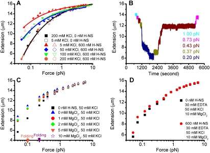
Magnesium-dependent binding modes of H-NS. (A) Force extension curve of DNA in the absence of magnesium. Two independent data sets are shown at each KCl concentration. The solid and open squares are the reference curves of DNA in buffers alone. The DNA becomes less stiff (less extended at the respective forces) as the KCl concentration increases. (B) Time course of DNA folding/unfolding in the presence of 50 mM KCl, 10 mM MgCl2, and 600 nM H-NS. Folding occurred at 0.2 pN and unfolding occurred at 0.43 pN. Complete unfolding occurred at 0.73 pN. It is a representative example of five experiments that were performed, in which folding occurred under 0.2–0.3 pN of force and subsequent unfolding occurred under 0.4–1 pN. (C) Switching H-NS from stiffening to bridging mode by increasing the concentration of MgCl2. Apparent stiffening occurred at ≤5 mM MgCl2. Folding (bridging) occurred at ≥5 mM MgCl2. The orange arrow indicates folding at ∼0.1 pN in the presence of 5 mM MgCl2. This bridged DNA was then completely unfolded at a larger force of ∼7 pN, during which the MgCl2 concentration was increased to 10 mM. The force was then incrementally reduced in the 10 mM MgCl2 buffer (purple stars), and bridging occurred at a higher stretching force (>0.2 pN; purple arrow). Stiffening (>0.1 pN) and folding (≤0.1 pN) coexist at 5 mM MgCl2. The H-NS concentration was fixed at 600 nM. (D) Effect of magnesium chelation by EDTA. The resulting extension of DNA in the presence of H-NS (red solid circles) is longer than that in the absence of H-NS (black squares), indicating that the DNA becomes stiffer. No folding was observed in the force range applied.
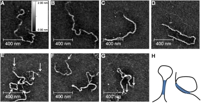
(A–D) AFM imaging of DNA–H-NS complexes under stiffening conditions. The buffer (pH 7.4) contained 10 mM Tris, 5 mM KCl, 0 mM MgCl2, and 600 nM H-NS. (A,B) Incubated for 40 min. (C,D) Incubated for 4 h. The brighter regions indicate the H-NS-bound regions, while the darker regions indicate the naked DNA backbone. (E–G) Imaging of DNA–H-NS complexes under bridging conditions. The buffer (pH 7.4) contained 10 mM Tris, 50 mM KCl, 10 mM MgCl2, and 600 nM H-NS. Incubation time was fixed at 40 min. Large linear hairpin forms and circular forms were observed. (H) Two boundary conformations of the seeding loop: almost anti-parallel (left), and almost parallel (right).
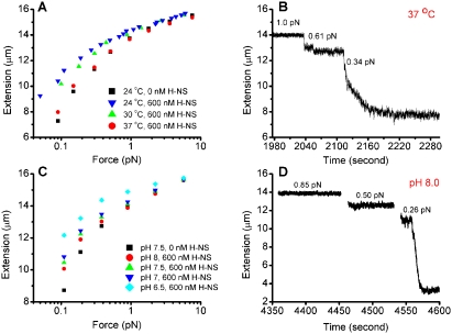
Susceptibility to temperature (A,B) and pH (C,D) under stiffening conditions (50 mM KCl and 0 mM MgCl2) and under bridging conditions (50 mM KCl and 10 mM MgCl2). (A) The stiffening effect decreases as the temperature increases. (B) The bridging mode was not affected by changing the temperature. At 37°C, folding still occurred at ∼0.3 pN. (C) Stiffening decreases with alkaline pH. (D) Bridging was unaffected by changing the pH in the same range; i.e., folding still occurred at ∼0.3 pN at pH 8.
H-NS interconverts between bridging and stiffening modes without being released from DNA. (A) Switching from stiffened DNA in 600 nM H-NS, 5 mM KCl, and 0 mM MgCl2 to bridging buffer in the absence of H-NS (0 mM H-NS, 50 mM KCl, 10 mM MgCl2) resulting in bridging. The blue arrow on the force axis indicates the force where the folding occurred. (B) Switching from bridged DNA in 600 nM H-NS, 50 mM KCl, and 10 mM MgCl2 to stiffening buffer in the absence of H-NS (0 mM H-NS, 5 mM KCl, 0 mM MgCl2) resulted in stiffening.
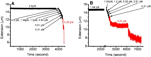
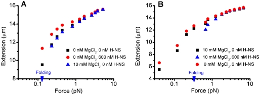
Precoated DNA can still fold under bridging buffer conditions (pH 7.4, 10 mM MgCl2, 50 mM KCl). (A) At 600 nM H-NS, DNA was kept in an extended conformation under 2.6 pN for ∼1 h. When the force was subsequently decreased to ∼0.25 pN, folding occurred (in red). (B) A similar experiment as in A was performed at 2.4 μM H-NS. Again, folding occurred when the force decreased to ∼0.25 pN (in red). Compared with the folding observed in the presence of 600 nM H-NS, the folding speed in 2.4 μM H-NS was reduced by >30-fold.
References
-
- Atlung T, Ingmer H. H-NS: A modulator of environmentally regulated gene expression. Mol Microbiol. 1997;24:7–17. - PubMed
-
- Bloch V, Yang YS, Margeat E, Chavanieu A, Auge MT, Robert B, Arold S, Rimsky S, Kochoyan M. The H-NS dimerization domain defines a new fold contributing to DNA recognition. Nat Struct Biol. 2003;10:212–218. - PubMed
-
- Bouffartigues E, Buckle M, Badaut C, Travers A, Rimsky S. H-NS cooperative binding to high-affinity sites in a regulatory element results in transcriptional silencing. Nat Struct Mol Biol. 2007;14:441–448. - PubMed
Publication types
MeSH terms
Substances
Grants and funding
LinkOut - more resources
Full Text Sources
Other Literature Sources
Miscellaneous