. 2010 Feb 15;24(4):411-22.
doi: 10.1101/gad.1878110.

A highly coordinated cell wall degradation machine governs spore morphogenesis in Bacillus subtilis

Affiliations

A highly coordinated cell wall degradation machine governs spore morphogenesis in Bacillus subtilis

Cecile Morlot et al. Genes Dev. .

Abstract

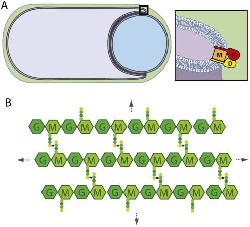
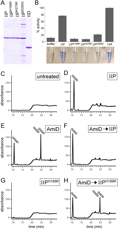
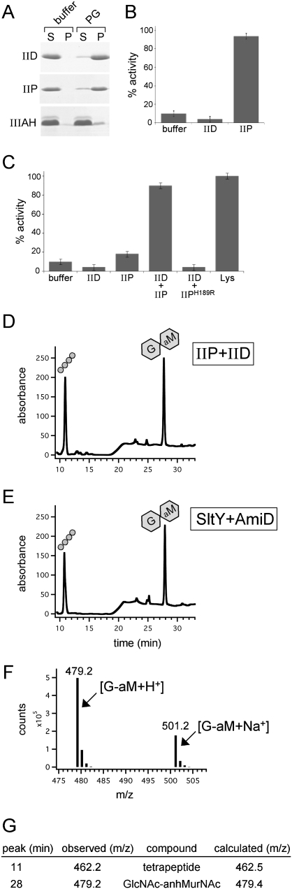
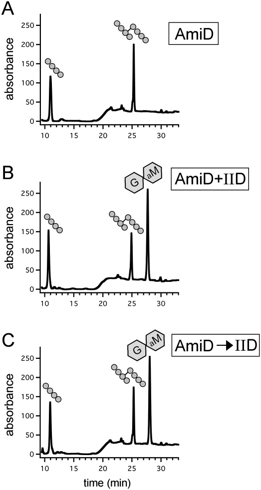
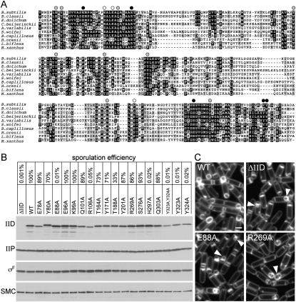
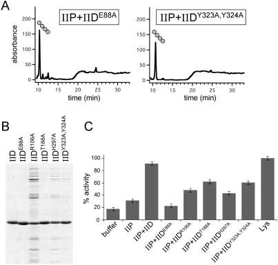
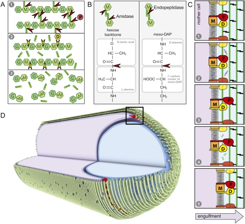
How proteins catalyze morphogenesis is an outstanding question in developmental biology. In bacteria, morphogenesis is intimately linked to remodeling the cell wall exoskeleton. Here, we investigate the mechanisms by which the mother cell engulfs the prospective spore during sporulation in Bacillus subtilis. A membrane-anchored protein complex containing two cell wall hydrolases plays a central role in this morphological process. We demonstrate that one of the proteins (SpoIIP) has both amidase and endopeptidase activities, such that it removes the stem peptides from the cell wall and cleaves the cross-links between them. We further show that the other protein (SpoIID) is the founding member of a new family of lytic transglycosylases that degrades the glycan strands of the peptidoglycan into disaccharide units. Importantly, we show that SpoIID binds the cell wall, but will only cleave the glycan strands after the stem peptides have been removed. Finally, we demonstrate that SpoIID also functions as an enhancer of SpoIIP activity. Thus, this membrane-anchored enzyme complex is endowed with complementary, sequential, and stimulatory activities. These activities provide a mechanism for processive cell wall degradation, supporting a model in which circumferentially distributed degradation machines function as motors pulling the mother cell membranes around the forespore.


Figures

Figure 1.
Figure 1.
Schematic diagram of the engulfment complex. (A) A sporangium during the morphological processes of engulfment. The membranes of the mother cell (purple) are migrating around the forespore (blue). The cell wall PG encasing the two cells is in green. IID (D), IIP (P), and IIM (M) are all made in the mother cell and localize to the leading edge of the engulfing septal membrane. IID (D) and IIP (P) both have a single N-terminal transmembrane segment and a large extracellular domain. IIM (M) is predicted to have five transmembrane segments. Genetic, biochemical, and cytological analysis indicate that these three proteins reside in a membrane complex (Aung et al. 2007; Chastanet and Losick 2007). (B) Schematic diagram of the PG meshwork. Glycan chains (hexagons) composed of GlcNAc (G) and MurNAc (M) are linked by glycosidic bonds. Attached to the MurNAc (M) sugars are short peptides (balls) that cross-link adjacent glycan strands, generating a continuous 3D meshwork that envelops the bacterium. Gram-positive bacteria like B. subtilis have multiple layers of PG.
Figure 2.
Figure 2.
IIP has both amidase and endopeptidase activity. (A) Coomassie-stained gel of purified proteins. The extracellular domains of wild-type IID, wild-type IIP, and three IIP mutants (Chastanet and Losick 2007) are shown. (B) Cell wall-degrading activities of IIP and IIP mutants using the RBB dye release assay. All reactions contained 4 μM protein. The dye-coupled soluble products released by the IIP proteins for 30 min at 37°C were normalized to the release by 4 μM lysozyme (Lys). Lysozyme cleaves the glycan strands, yielding soluble disaccharide peptide products. Similarly, amidases like AmiD release soluble glycan chains by destroying peptide cross-bridges (Supplemental Fig. S1). The supernatants after incubation with the dye-coupled PG are shown below the histograms. (C–H) RP-HPLC elution profiles of the soluble products after treatment of unlabeled PG with indicated proteins. (C) No treatment of the PG. (D) Incubation with 4 μM IIP. (E) Incubation with 4 μM E. coli AmiD. (F) Incubation with 4 μM AmiD followed by heat inactivation and incubation with 4 μM IIP. Note the loss of cross-linked tetrapeptide after incubation with IIP. (G) Incubation with 4 μM IIPH189R. (H) Incubation with 4 μM AmiD followed by heat inactivation and incubation with 4 μM IIPH189R. Tetrapeptide and cross-linked tetrapeptide products are shown schematically above the elution peaks.
Figure 3.
Figure 3.
IID activity requires IIP. (A) IID binds PG. The extracellular domains of IID, IIP, and a control protein that does not bind PG (IIIAH) were incubated separately with buffer or E. coli PG. Coomassie-stained gel of the supernatant (S) and pellet (P) fractions after a 16,000g spin is shown. (B) IID has no cell wall-degrading activity on its own. Analysis of IID using the dye release assay. All reactions contained 4 μM protein. The activities of IIP and IID were normalized to lysozyme (data not shown). (C) Analysis of mixtures of IID and IIP using the dye release assay. All reactions with IID and IIP contained 1 μM each protein. The lysozyme reaction contained 4 μM protein. (D) RP-HPLC elution profiles of the soluble products after treatment of unlabeled PG with IIP and IID. The tetrapeptide (balls) and anhydrodisaccharide (hexagons) products are shown schematically above the elution peaks. (E) RP-HPLC elution profiles of the soluble products after treatment of unlabeled PG with SltY and AmiD (see the Materials and Methods). (F) LC/MS analysis in positive ion mode of the product in the 28-min fraction from D. Shown are the mass to charge ratios (m/z) of the ions detected and their relative abundance. (G) Table of calculated and observed m/z of the products in fractions 11 and 28 from D. The product in fraction 11 had an m/z consistent with a tetrapeptide. The product in fraction 28 had an m/z consistent with GlcNAc–anhydro MurNAc.
Figure 4.
Figure 4.
IID cleaves naked glycan strands. RP-HPLC elution profiles of the soluble products after treatment of unlabeled PG with the indicated proteins (4 μM each). (A) Incubation with AmiD. (B) Incubation with AmiD and IID. (C) Incubation with AmiD followed by heat inactivation and treatment with IID. The tetrapeptide, cross-linked tetrapeptide, and anhydrodisaccharide products are shown schematically above the elution peaks.
Figure 5.
Figure 5.
Identification of amino acid residues in IID that are required for function. (A) Amino acid sequence alignment of the extracellular domains of IID homologs from representative phyla. Bacteria include B. subtilis, Bacillus clausii KSM-K16, Eubacterium dolichum DSM 3991, Clostridium beijerinckii NCIMB 8052, Anabaena variabilis ATCC 29413, Syntrophomonas wolfei, Bacteroides capillosus ATCC 29799, Halothermothrix orenii H 168, Leptospira biflexa, and Myxococcus xanthus. See Supplemental Figure S6 for a larger alignment. Conserved amino acids (black boxes) and similar residues (gray boxes) are highlighted. Amino acid substitutions that were tested are indicated above the sequence. Black circles indicate a strong block in engulfment and a >1000-fold defect in sporulation efficiency. Gray circles indicate aberrant engulfment as assessed by fluorescence microscopy. White circles indicate alanine substitutions with no significant affect on engulfment or sporulation efficiency. The first amino acid residue shown for B. subtilis IID is amino acid 72. (B) Sporulation efficiencies of B. subtilis strains containing wild-type IID, a IID-null mutant (ΔIID), and IID mutants with the indicated amino acid substitutions. Cells from the same strains were collected at hour 2 of sporulation, and IID and IIP protein levels were analyzed by immunoblot. All strains efficiently entered sporulation, as judged by the levels of the sporulation transcription factor σF. The SMC protein was used to control for loading. (C) At hour 2 of sporulation, the indicated strains were analyzed by fluorescence microscopy using the membrane dye TMA-DPH. The bulged septal membranes in cells lacking IID or those producing IIDE88A (E88A) are indicated (yellow carets). Impaired engulfment due to the IIDR269A (R269A) mutation is indicated with the white caret. Supplemental Figure S8 shows fluorescence images of all 17 mutants. Bar, 1 μm.
Figure 6.
Figure 6.
IID enhances IIP activity. (A) RP-HPLC elution profiles of the soluble products after treatment of unlabeled PG with IIP and the indicated IID mutants. The tetrapeptide product is shown schematically. (B) Coomassie-stained gel of purified proteins. The extracellular domains of wild-type IID and five IID mutants are shown. (C) Analysis of IIP activity in the presence of the IID mutants using the RBB dye release assay. All reactions with IID and IIP contained 1 μM each protein. The lysozyme reaction contained 4 μM protein.
Figure 7.
Figure 7.
Coordinated activities in the engulfment complex drive membrane migration around the forespore. (A) Schematic representation of the complementary and sequential activities of IIP and IID. (1) In the diagram, IIP cleaves the stem peptide from the glycan strand and the cross-links between the tetrapeptides. (2) Following peptide cleavage by IIP, IID cleaves the denuded glycan strands into anhydrodisaccharides. (B) Comparison of the amide bonds cleaved by IIP. (C) Schematic diagram of the proposed catalytic cycle of the engulfment complex. (1) IIP (P) and IID (D) in complex with IIM (M) bind the PG. (2) IID stimulates the amidase activity of IIP, resulting in cleavage of the stem peptides and peptide cross-links (black). (3) IIP is released from the PG and rebinds at a nearby site. (4) IID cleaves the denuded glycan strands (green). (5) IID is released from the PG and rebinds adjacent to IIP. The leading edge of the engulfing mother cell membrane (dark purple) moves toward the forespore pole (to the right). (D) Circumferentially distributed engulfment complexes drive membrane movement around the forespore. Shown is a schematic diagram of a sporangium during the morphological processes of engulfment. The membranes of the mother cell (purple) are migrating around the forespore (blue). The glycan strands (green) are shown running perpendicular to the long axis of the cell, as has been proposed for E. coli and C. crescentus (Holtje 1998; Gan et al. 2008). The hoops of glycan strands are stitched together by the cross-linked tetrapeptides. IID (yellow) and IIP (red) are anchored in the leading edge of the mother cell membrane. Processive degradation of the PG drives the mother cell membranes toward the cell pole. For simplicity, we show the engulfment complex acting on the basement layer of the PG meshwork. It is possible that the complex degrades more than one layer, and not necessarily the layer adjacent to the forespore.

Similar articles

Cited by

References

    1. Abanes-De Mello A, Sun YL, Aung S, Pogliano K. A cytoskeleton-like role for the bacterial cell wall during engulfment of the Bacillus subtilis forespore. Genes & Dev. 2002;16:3253–3264. - PMC - PubMed
    1. Altschul SF, Madden TL, Schaffer AA, Zhang J, Zhang Z, Miller W, Lipman DJ. Gapped BLAST and PSI-BLAST: A new generation of protein database search programs. Nucleic Acids Res. 1997;25:3389–3402. - PMC - PubMed
    1. Aung S, Shum J, Abanes-De Mello A, Broder DH, Fredlund-Gutierrez J, Chiba S, Pogliano K. Dual localization pathways for the engulfment proteins during Bacillus subtilis sporulation. Mol Microbiol. 2007;65:1534–1546. - PMC - PubMed
    1. Bendezu FO, Hale CA, Bernhardt TG, de Boer PA. RodZ (YfgA) is required for proper assembly of the MreB actin cytoskeleton and cell shape in E. coli. EMBO J. 2009;28:193–204. - PMC - PubMed
    1. Blackburn NT, Clarke AJ. Identification of four families of peptidoglycan lytic transglycosylases. J Mol Evol. 2001;52:78–84. - PubMed

Publication types

MeSH terms

LinkOut - more resources