. 2010 Apr 16;285(16):12210-22.
doi: 10.1074/jbc.M109.062364. Epub 2010 Feb 16.

Identification of a linear epitope in sortilin that partakes in pro-neurotrophin binding

Affiliations

Identification of a linear epitope in sortilin that partakes in pro-neurotrophin binding

Olga Serup Andersen et al. J Biol Chem. .

Abstract

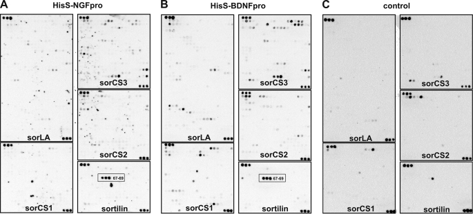
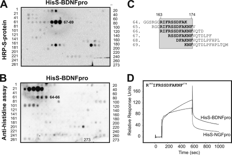
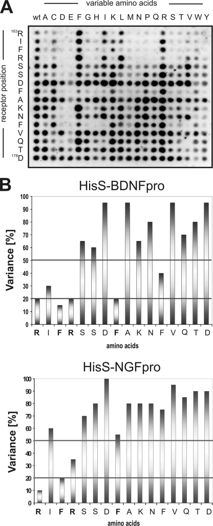
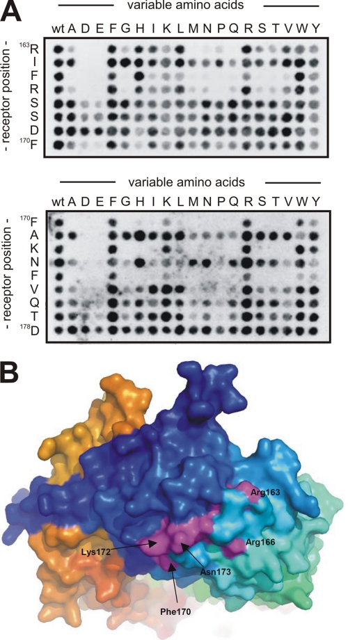
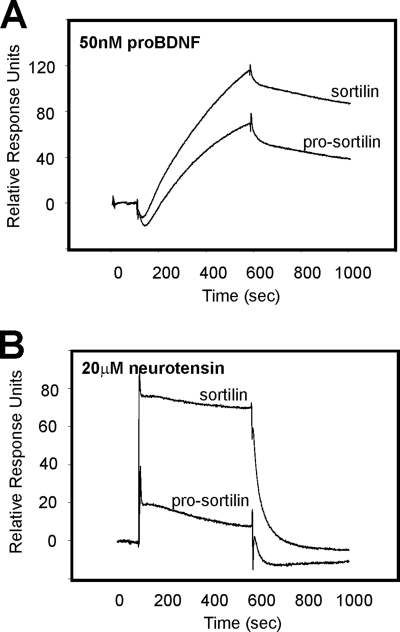
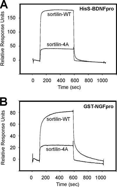
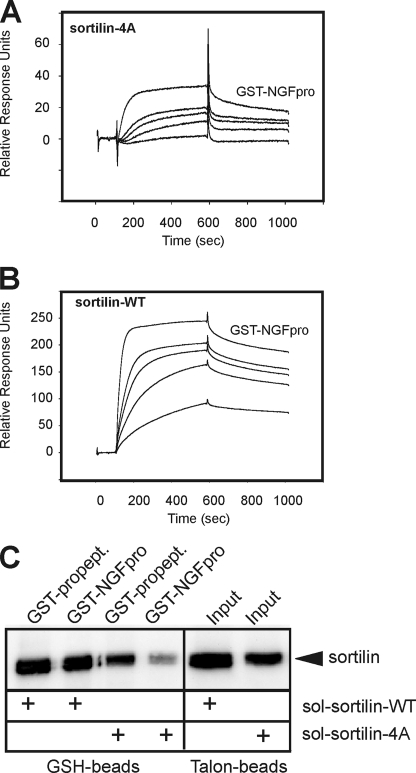
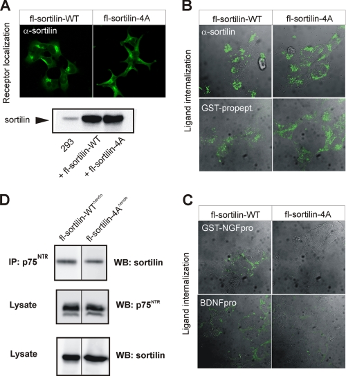
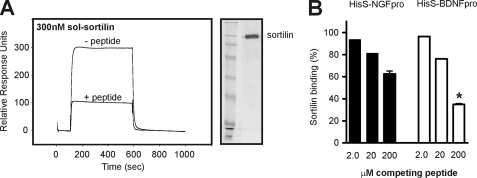
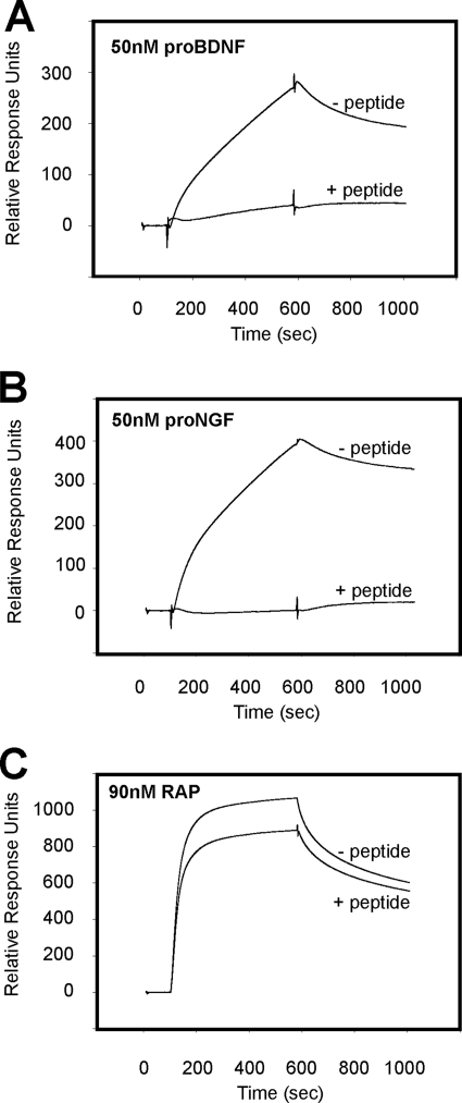
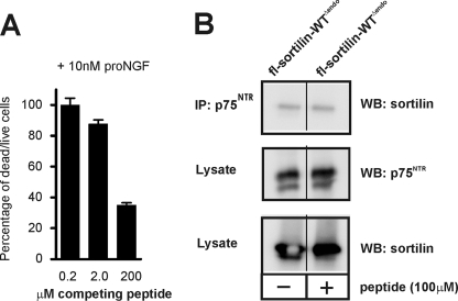
Sortilin acts as a cell surface receptor for pro-neurotrophins (pro-NT) that upon complex formation with the p75 neurotrophin receptor (p75(NTR)) is able to signal neuronal cell death. Here we screened a sortilin peptide library comprising 16-mer overlapping sequences for binding of the pro-domains of nerve growth factor and brain-derived neurotrophic factor. We find that a linear surface-exposed sequence, (163)RIFRSSDFAKNF(174), constitutes an important pro-NT binding epitope in sortilin. Systematic mutational analysis revealed residues Arg(163), Phe(165), Arg(166), and Phe(170) to be critical for the interaction. Expression of a sortilin mutant in which these four amino acids were substituted by alanines disrupted pro-NT binding without affecting receptor heterodimerization with p75(NTR) or binding of ligands that selectively engages the centrally located tunnel in the beta-propeller of sortilin. We furthermore demonstrate that a peptide comprising the ligand-binding epitope can prevent pro-NT-induced apoptosis in RN22 schwannoma cells.


Figures

FIGURE 1.
FIGURE 1.
Analysis of the NGF and the BDNF pro-domain binding to the mammalian VPS10p receptors by SPOT analysis. A peptide library containing a total of 2181 peptides represented by 734 from sorLA, 403 from sorCS3, 389 from sorCS1, 382 from sorCS2, and 273 from sortilin as overlapping 16-mer peptides of the five human VPS10p-domain-containing receptors sortilin (CAA66904, 831 aa), sorLA (AAC50891, 2214 aa), sorCS1(AAM43811, 1179 aa), sorCS2 (Q96PQ0, 1159 aa), and sorCS3(Q9UPU3, 1222 aa). The membrane was incubated either in the presence of 10 μg/ml HisS-NGFpro (A), 10 μg/ml HisS-BDNFpro (B), or in the absence of ligand (C). Detection was carried out by incubation of the membrane with an HRP-conjugate of S-protein that recognizes the S-peptide tag of bound NT pro-domains. Each receptor library is cornered at the upper left and lower right corner by three control peptides equivalent to the S-peptide tag (amino acid sequence KETAAAKFERQHMS). The specific binding site for both HisS-NGFpro and HisS-BDNFpro (A and B), which is not seen for the control experiment in absence of ligand (C), is indicated for the three sortilin peptides (SPOT numbers 67–69; red box).
FIGURE 2.
FIGURE 2.
Identification of a linear pro-NT binding site within sortilin. A and B, sortilin represented as 273 overlapping peptides generated by SPOT synthesis on a cellulose membrane with peptides 64–69 binding to HisS-BDNFpro, as detected by either HRP-S-protein (A, SPOT numbers 67–69) or antibody immunoassay against the polyhistidine tag (B, SPOT numbers 64–66). C, peptide sequences of the six peptides 64–69 found to interact with the pro-domains of BDNF and NGF. Residues corresponding to the sortilin fragment 163–174 are boxed. D, SPR analysis of the 12-mer sortilin-derived peptide (residues 163RIFRSSDFAKNF174) binding to flow cells with immobilized HisS-BDNFpro and HisS-NGFpro.
FIGURE 3.
FIGURE 3.
Substitution analysis of the linear sortilin sequence. A, a representative substitutional binding analysis of HisS-BDNFpro to peptides with the wild-type sortilin sequence 163RIFRSSDFAKNFVQTD178 listed to the left on the membrane and detection using the anti-histidine immunoassay. B, binding assays as demonstrated in panel A were carried out for both HisS-BDNFpro and HisS-NGFpro, and signal intensities, measured in Boehringer Light Units (BLU), were quantified for each peptide. The percentage of replacement variability (V) of each sequence position have been calculated (V = BLU/20 × 100%) and plotted against each amino acid within the peptide (residues 163–178).
FIGURE 4.
FIGURE 4.
Mutational binding analysis of HisS-pro-BDNF to short sortilin fragments. A, HisS-BDNFpro binding analysis to peptides with the wild-type sortilin sequences 163RIFRSSDF170 and 170FAKNFVQTD178 listed to the left on each membrane. Binding to mutant peptides where each amino acid has been substituted with the 20 naturally occurring amino acids is used for identification of specific residues important for interaction with the immature part of pro-BDNF. The substitution analysis clearly identifies the two sequences 163RIFR166 (upper part) and 170FAKNF174 (lower part) as specific binding sites for HisS-BDNFpro. B, crystal structure presentation of sortilin showing the surface exposure of side chains from several identified key residues in purple (e.g. Arg163, Arg166, Phe170, and Lys172). Structure was drawn from coordinates present in the Protein Data Bank with accession code 3F6K.
FIGURE 5.
FIGURE 5.
Differential binding of pro-BDNF and neurotension to sortilin and pro-sortilin. A, SPR analysis showing that unprocessed pro-BDNF (50 nm) binds nearly as efficiently to the receptor in the presence (pro-sortilin) as in the absence (sortilin) of the receptor propeptide. Pro-sortilin is a receptor variant unable to cleave off the propeptide due to mutation of the furin recognition site. B, binding of neurotensin (20 μm) is strongly ablated for pro-sortilin as compared with binding to sortilin without the presence of its propeptide.
FIGURE 6.
FIGURE 6.
Mutation of the linear binding site specifically impairs binding of both the NGF and the BDNF pro-domains. SPR analysis showing reduced binding of equal amounts (analyte concentration: 200 nm) of the soluble extracellular domains of sortilin-4A compared with sortilin-WT when binding is tested to both the immobilized BDNF pro-domain (HisS-BDNFpro, A) and the immobilized NGF pro-domain (GST-NGFpro, B).
FIGURE 7.
FIGURE 7.
GST-NGFpro binding to sortilin-WT and sortilin-4A. SPR analysis showing concentration series of GST-NGFpro (10, 20, 30, 40, and 50 nm) tested for binding to immobilized extracellular domains of sortilin-4A (A) and sortilin-WT (B), demonstrating a strong decrease in the binding capacity for the NGF pro-domain upon the quadruple mutation in sortilin-4A. A higher than 10-fold decrease in affinity (WT: KD ∼2 nm versus 4A: KD ∼26 nm) was estimated using the BIAevaluation software. C, medium from EBNA 293 cells producing either the soluble ectodomain of sortilin-WT or sortilin-4A were incubated with a GST-tagged variant of the receptor propeptide (GST-propept) or GST-NGFpro and precipitated by glutathione (GSH) beads. The amount of total secreted sortilin-WT and sortilin-4A (i.e. input) was determined by precipitation using Talon beads binding to the histidine tag within the sortilin domains. The precipitated proteins were subjected to SDS-PAGE analysis and visualized by Western blot analysis for sortilin.
FIGURE 8.
FIGURE 8.
Decreased binding of pro-NT to sortilin-4A within cells. A, immunostaining of HEK 293 cells transfected with constructs for full-length (fl)-sortilin-WT or sortilin-4A with an antibody against sortilin. Protein expression levels tested by Western blot analysis of cell lysates. B and C, cells were labeled with ligands at 4 °C followed by endocytosis for 30 min at 37 °C before fixation and internalized ligand visualization by staining. Ligands applied were either a sortilin antibody (against the receptor extracellular domain; α-sortilin), the receptor propeptide (GST-propept) (B), GST-NGFpro, or BDNFpro (C). D, cells co-transfected with constructs for sortilin variants devoid of endocytosis (fl-sortilin-WTΔendo or fl-sortilin-4AΔendo) together with p75NTR were immunoprecipitation using an antibody against p75NTR. Lysates and precipitated proteins were subjected to SDS-PAGE and analyzed by Western blotting for either p75NTR or sortilin as indicated.
FIGURE 9.
FIGURE 9.
Sortilin-derived peptide specifically competes binding of pro-NT to sortilin. A, recombinant sortilin was purified from 293 cells (indicated to the right by silver-stained SDS-PAGE analysis), and used for SPR studies to immobilized HisS-BDNFpro. The signal of 300 RU observed for binding in the absence of the peptide sort166–181 (− peptide) was significantly lowered to 100 RU in the presence of 200 μm linear sortilin antagonist (+ peptide). B, sortilin binding to either HisS-NGFpro or HisS-BDNFpro was determined by SRP analysis (as exemplified in A) and the inhibition by increasing amounts of sort166–181 (at 2, 20, and 200 μm) was plotted relative to the observed interaction in the absence of competitor (in percentage). Values represent the mean ± S.E. from three experiments. *, p = 0.0007; two-tailed Student's t test.
FIGURE 10.
FIGURE 10.
Selective competition of ligands by sortilin-derived peptide antagonist. SPR binding analysis of 50 nm unprocessed pro-BDNF (A), 50 nm unprocessed pro-NGF (B), and 90 nm RAP (C) to immobilized sortilin in the absence and presence of the sort166–181 peptide (100 μm). Specific inhibition of pro-NGF and pro-BDNF is observed, whereas the interaction between sortilin and RAP is largely intact.
FIGURE 11.
FIGURE 11.
A sortilin-derived peptide blocks pro-NGF-induced cell death. A, RN22 schwannoma cells were incubated in the presence of 10 nm pro-NGF and increasing concentrations (0.2, 2.0, and 200 μm) of the sort166–181 peptide. The amount of pro-NGF-induced cell death after 72 h were quantified using a fluorescence-based assay, and plotted against competitor concentrations as the ratio between dead and live cells in percentage. B, cells co-transfected with constructs for a sortilin variant devoid of endocytosis (fl-sortilin-WTΔendo) together with p75NTR were incubated in the absence or presence of 100 μm sort166–181 peptide, and immunoprecipitated using an antibody against p75NTR. Lysates and precipitated proteins were subjected to SDS-PAGE and analyzed by Western blotting for either p75NTR or sortilin as indicated.

Similar articles

Cited by

References

    1. Willnow T. E., Petersen C. M., Nykjaer A. (2008) Nat. Rev. Neurosci. 9, 899–909 - PubMed
    1. Andersen O. M., Reiche J., Schmidt V., Gotthardt M., Spoelgen R., Behlke J., von Arnim C. A., Breiderhoff T., Jansen P., Wu X., Bales K. R., Cappai R., Masters C. L., Gliemann J., Mufson E. J., Hyman B. T., Paul S. M., Nykjaer A., Willnow T. E. (2005) Proc. Natl. Acad. Sci. U.S.A. 102, 13461–13466 - PMC - PubMed
    1. Rogaeva E., Meng Y., Lee J. H., Gu Y., Kawarai T., Zou F., Katayama T., Baldwin C. T., Cheng R., Hasegawa H., Chen F., Shibata N., Lunetta K. L., Pardossi-Piquard R., Bohm C., Wakutani Y., Cupples L. A., Cuenco K. T., Green R. C., Pinessi L., Rainero I., Sorbi S., Bruni A., Duara R., Friedland R. P., Inzelberg R., Hampe W., Bujo H., Song Y. Q., Andersen O. M., Willnow T. E., Graff-Radford N., Petersen R. C., Dickson D., Der S. D., Fraser P. E., Schmitt-Ulms G., Younkin S., Mayeux R., Farrer L. A., St George-Hyslop P. (2007) Nat. Genet. 39, 168–177 - PMC - PubMed
    1. Scherzer C. R., Offe K., Gearing M., Rees H. D., Fang G., Heilman C. J., Schaller C., Levey A. I., Lah J. J. (2004) Arch. Neurol. 61, 1200–1205 - PubMed
    1. Clee S. M., Yandell B. S., Schueler K. M., Rabaglia M. E., Richards O. C., Raines S. M., Kabara E. A., Klass D. M., Mui E. T., Stapleton D. S., Gray-Keller M. P., Young M. B., Stoehr J. P., Lan H., Boronenkov I., Raess P. W., Flowers M. T., Attie A. D. (2006) Nat. Genet. 38, 688–693 - PubMed

Publication types

MeSH terms