. 2010 Apr 16;285(16):12378-89.
doi: 10.1074/jbc.M109.093583. Epub 2010 Feb 16.

Deciphering the catalytic domain of colicin M, a peptidoglycan lipid II-degrading enzyme

Affiliations

Deciphering the catalytic domain of colicin M, a peptidoglycan lipid II-degrading enzyme

Hélène Barreteau et al. J Biol Chem. .

Abstract

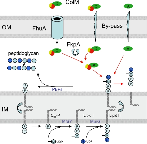
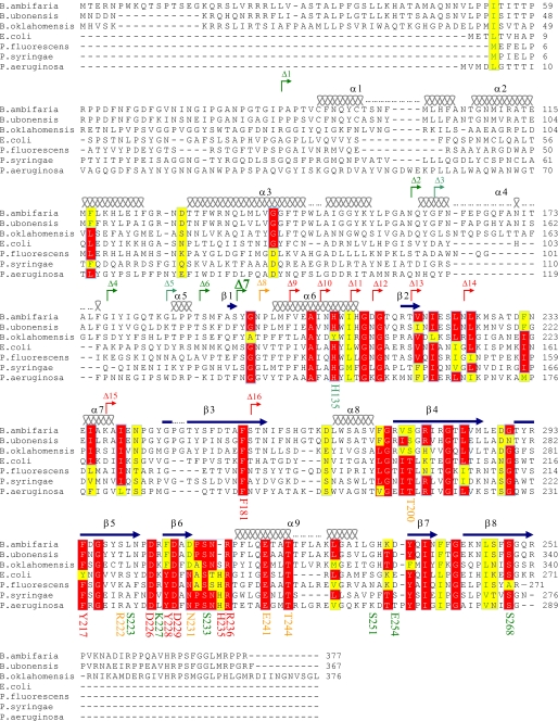
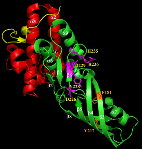
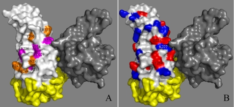
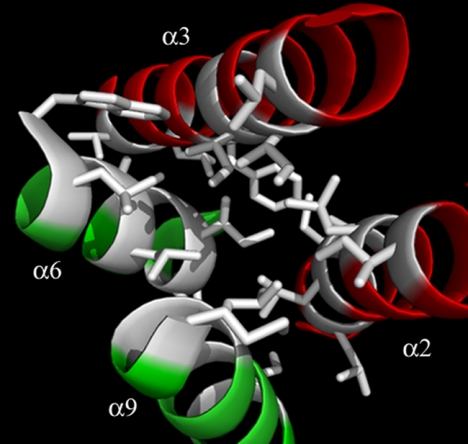
Colicin M inhibits Escherichia coli peptidoglycan synthesis through cleavage of its lipid-linked precursors. It has a compact structure, whereas other related toxins are organized in three independent domains, each devoted to a particular function: translocation through the outer membrane, receptor binding, and toxicity, from the N to the C termini, respectively. To establish whether colicin M displays such an organization despite its structural characteristics, protein dissection experiments were performed, which allowed us to delineate an independent toxicity domain encompassing exactly the C-terminal region conserved among colicin M-like proteins and covering about half of colicin M (residues 124-271). Surprisingly, the in vitro activity of the isolated domain was 45-fold higher than that of the full-length protein, suggesting a mechanism by which the toxicity of this domain is revealed following primary protein maturation. In vivo, the isolated toxicity domain appeared as toxic as the full-length protein under conditions where the reception and translocation steps were by-passed. Contrary to the full-length colicin M, the isolated domain did not require the presence of the periplasmic FkpA protein to be toxic under these conditions, demonstrating that FkpA is involved in the maturation process. Mutational analysis further identified five residues that are essential for cytotoxicity as well as in vitro lipid II-degrading activity: Asp-229, His-235, Asp-226, Tyr-228, and Arg-236. Most of these residues are surface-exposed and located relatively close to each other, hence suggesting they belong to the colicin M active site.


Figures

FIGURE 1.
FIGURE 1.
Synthesis of peptidoglycan lipid-linked intermediates and mode of action of ColM. The inner (IM) and outer (OM) membranes are depicted by the gray boxes. The MraY enzyme catalyzes the transfer of the phospho-MurNAc-pentapeptide from the nucleotide precursor onto the carrier lipid undecaprenyl phosphate (C55-P), yielding lipid I; subsequently, the MurG enzyme adds the GlcNAc moiety, yielding lipid II, which is translocated from the inner side of the membrane to the outer side (38, 39). Thereafter, the disaccharide-pentapeptide motif is polymerized and incorporated into the peptidoglycan through the action of the penicillin-binding proteins (PBPs) releasing the lipid carrier in a pyrophosphate form that will be recycled (40, 41). Incoming ColM was shown to cleave the lipid-linked intermediates between the C55 and pyrophosphoryl-disaccharide-peptide motifs (6). In the present report, an independent ColM toxicity domain (green) was identified (residues 124–271), which was active in vivo under conditions where the FhuA receptor was by-passed. In contrast to the full-length ColM, the independent toxicity domain did not require the periplasmic FkpA protein to be toxic. These data suggest that ColM exists in two active (A) and inactive (I) conformations, FkpA being involved in the activation process. The reception and translocation domains of ColM are represented in red and yellow, respectively.
FIGURE 2.
FIGURE 2.
Sequence alignment of ColM with its homologues. Amino acid sequences of E. coli ColM (accession number P05820) and its homologues from Burkholderia ambifaria MC40–6, Burkholderia ubonensis Bu, Burkholderia oklahomensis Eo147, Pseudomonas fluorescens, Pseudomonas syringae pv. tomato DC3000 and Pseudomonas aeruginosa EXA13 were aligned using the ClustalW program. Based on the known three-dimensional structure of ColM (17), secondary structural elements are indicated at the top of the sequences, the wavy lines designating α-helices and the arrows designating β-strands. The numbers of these structural elements are given according to the structure. The conserved residues are color-coded in red for identical residues and yellow for similar ones. The starting points of each N-terminal deletion constructs (from Δ1 toward Δ16) are indicated by broken arrows. The truncations indicated in green exhibit high in vitro activities, whereas those in red had no activity (ColM-Δ8 had intermediate activity). The targeted residues for mutagenesis are also indicated by their respective numbers below the sequences. According to the ability of the corresponding mutants to inhibit cell growth, the residues were classified as non-important (green), important (orange), and essential residues (red).
FIGURE 3.
FIGURE 3.
Three-dimensional structure of ColM. Based together on structural data from Zeth et al. (17) and biochemical data from this study, the three functional domains are illustrated as follows: the translocation domain in yellow (residues 1–35), the receptor-binding domain in red (residues 36–123), and the toxicity domain in green (124–271). The residues whose substitution caused a total loss of cytotoxicity are indicated as sticks. Residues that likely belong to the active site (Asp-226, Tyr-228, Asp-229, His-235, and Arg-236) are indicated in magenta, and structurally important residues (Phe-181 and Tyr-217) are in orange. The figure was prepared with the atomic coordinates 3DA4 deposited by Zeth et al. (17) by using PyMOL (DeLano Scientific).
FIGURE 4.
FIGURE 4.
Surface representation of the ColM structure. Because ColM was crystallized as a dimer, one protomer is indicated in gray, the other protomer is illustrated as follows: the N-terminal and central domains (residues 1–123) are shown in yellow, and the toxicity domain is indicated in white. A, the data of the mutational analysis were mapped onto the structure of ColM. The amino acids whose mutation resulted in a total loss of cytotoxicity are in pink, and the amino acids with a less substantial effect in orange. B, ColM is in the same orientation as in panel A except the surface of the toxicity domain is colored on the basis of electrostatic potential, with negative charges in red and positive charges in blue.
FIGURE 5.
FIGURE 5.
Representation of the interface between the central and toxicity domains of ColM. The ribbon diagram of the α-helices from the central domain (α2 and α3, in red) and from the toxicity domain (α6 and α9, in green) is shown. This four-helix bundle forms a hydrophobic core constitutive of the interface between the two domains. The side chains of hydrophobic residues are shown as sticks.

Similar articles

Cited by

References

    1. Pugsley A. P. (1984) Microbiol. Sci. 1, 203–205 - PubMed
    1. Pugsley A. P. (1984) Microbiol. Sci. 1, 168–175 - PubMed
    1. Cascales E., Buchanan S. K., Duché D., Kleanthous C., Lloubès R., Postle K., Riley M., Slatin S., Cavard D. (2007) Microbiol. Mol. Biol. Rev. 71, 158–229 - PMC - PubMed
    1. Harkness R. E., Braun V. (1989) J. Biol. Chem. 264, 6177–6182 - PubMed
    1. Schaller K., Höltje J. V., Braun V. (1982) J. Bacteriol. 152, 994–1000 - PMC - PubMed

Publication types

MeSH terms

LinkOut - more resources