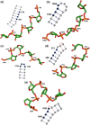
Arrangement of 3D structural motifs in ribosomal RNA
- PMID: 20159997
- PMCID: PMC2887949
- DOI: 10.1093/nar/gkq074
Arrangement of 3D structural motifs in ribosomal RNA
Abstract
Structural 3D motifs in RNA play an important role in the RNA stability and function. Previous studies have focused on the characterization and discovery of 3D motifs in RNA secondary and tertiary structures. However, statistical analyses of the distribution of 3D motifs along the RNA appear to be lacking. Herein, we present a novel strategy for evaluating the distribution of 3D motifs along the RNA chain and those motifs whose distributions are significantly non-random are identified. By applying it to the X-ray structure of the large ribosomal subunit from Haloarcula marismortui, helical motifs were found to cluster together along the chain and in the 3D structure, whereas the known tetraloops tend to be sequentially and spatially dispersed. That the distribution of key structural motifs such as tetraloops differ significantly from a random one suggests that our method could also be used to detect novel 3D motifs of any size in sufficiently long/large RNA structures. The motif distribution type can help in the prediction and design of 3D structures of large RNA molecules.
Figures


Similar articles
-
Finding 3D motifs in ribosomal RNA structures.Nucleic Acids Res. 2009 Mar;37(4):e29. doi: 10.1093/nar/gkn1044. Epub 2009 Jan 21. Nucleic Acids Res. 2009. PMID: 19158187 Free PMC article.
-
Sequence and structural conservation in RNA ribose zippers.J Mol Biol. 2002 Jul 12;320(3):455-74. doi: 10.1016/s0022-2836(02)00515-6. J Mol Biol. 2002. PMID: 12096903
-
RNA tertiary interactions in the large ribosomal subunit: the A-minor motif.Proc Natl Acad Sci U S A. 2001 Apr 24;98(9):4899-903. doi: 10.1073/pnas.081082398. Epub 2001 Apr 10. Proc Natl Acad Sci U S A. 2001. PMID: 11296253 Free PMC article.
-
Progress toward an understanding of the structure and enzymatic mechanism of the large ribosomal subunit.Cold Spring Harb Symp Quant Biol. 2001;66:33-42. doi: 10.1101/sqb.2001.66.33. Cold Spring Harb Symp Quant Biol. 2001. PMID: 12762006 Review. No abstract available.
-
The structural basis of large ribosomal subunit function.Annu Rev Biochem. 2003;72:813-50. doi: 10.1146/annurev.biochem.72.110601.135450. Annu Rev Biochem. 2003. PMID: 14527328 Review.
Cited by
-
Mining for recurrent long-range interactions in RNA structures reveals embedded hierarchies in network families.Nucleic Acids Res. 2018 May 4;46(8):3841-3851. doi: 10.1093/nar/gky197. Nucleic Acids Res. 2018. PMID: 29608773 Free PMC article.
-
GINClus: RNA structural motif clustering using graph isomorphism network.NAR Genom Bioinform. 2025 Apr 26;7(2):lqaf050. doi: 10.1093/nargab/lqaf050. eCollection 2025 Jun. NAR Genom Bioinform. 2025. PMID: 40290315 Free PMC article.
-
RNA structural motif recognition based on least-squares distance.RNA. 2013 Sep;19(9):1183-91. doi: 10.1261/rna.037648.112. Epub 2013 Jul 25. RNA. 2013. PMID: 23887146 Free PMC article.
-
RNAMotifScan: automatic identification of RNA structural motifs using secondary structural alignment.Nucleic Acids Res. 2010 Oct;38(18):e176. doi: 10.1093/nar/gkq672. Epub 2010 Aug 8. Nucleic Acids Res. 2010. PMID: 20696653 Free PMC article.
-
On the predictibility of A-minor motifs from their local contexts.RNA Biol. 2022 Jan;19(1):1208-1227. doi: 10.1080/15476286.2022.2144611. RNA Biol. 2022. PMID: 36384383 Free PMC article.
References
-
- Ahsen U, Schroeder R. RNA as a catalyst: natural and designed ribozymes. BioEssays. 2005;15:299–307. - PubMed
-
- Gilbert W. Origin of life: the RNA world. Nature. 1986;319:618.
-
- Ban N, Nissen P, Hansen J, Moore PB, Steitz TA. The complete atomic structure of the large ribosomal subunit at 2.4 Å resolution. Science. 2000;289:905–920. - PubMed

