

Protein kinase D1 inhibits cell proliferation through matrix metalloproteinase-2 and matrix metalloproteinase-9 secretion in prostate cancer
- PMID: 20160036
- PMCID: PMC3197700
- DOI: 10.1158/0008-5472.CAN-09-4155
Protein kinase D1 inhibits cell proliferation through matrix metalloproteinase-2 and matrix metalloproteinase-9 secretion in prostate cancer
Abstract
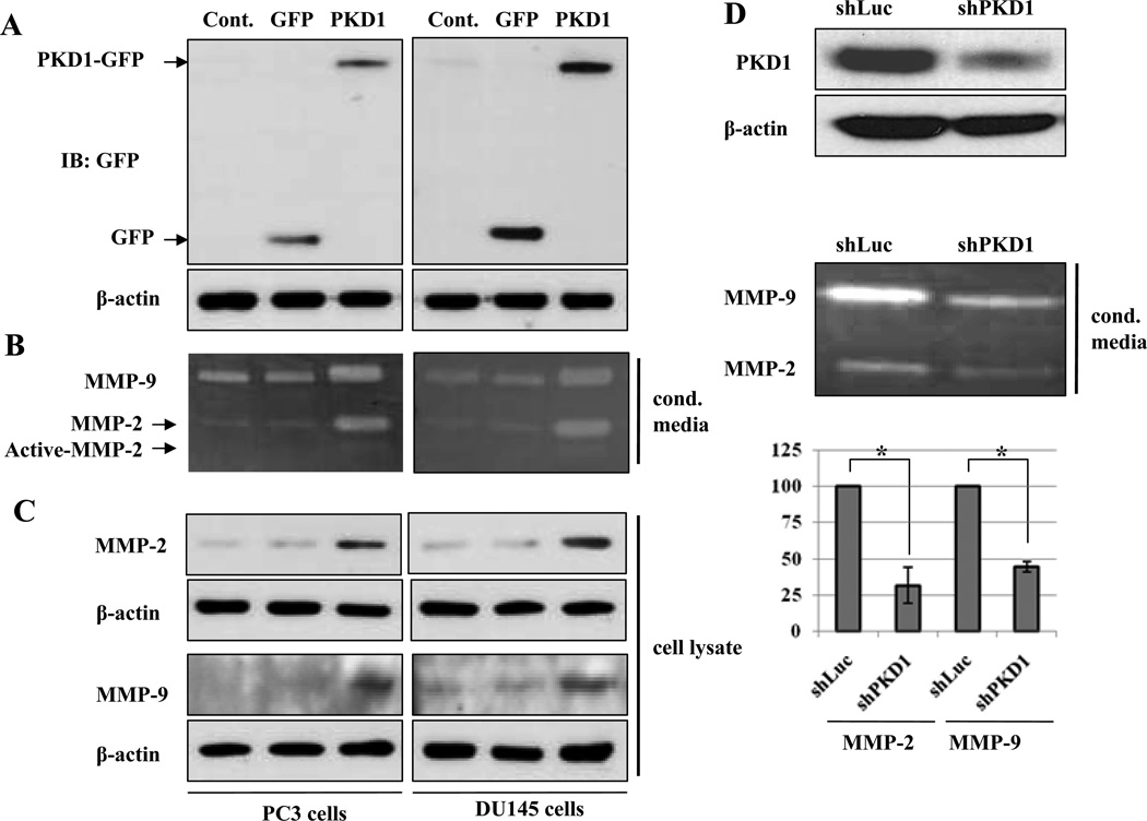
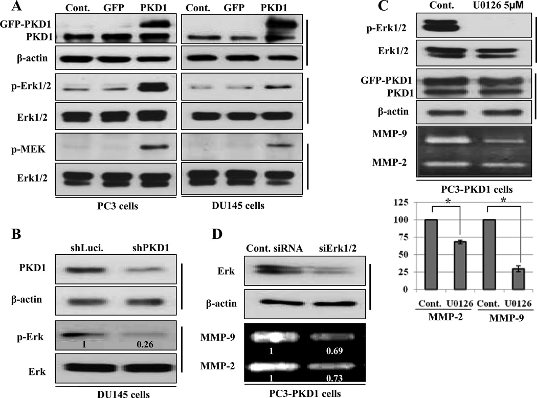
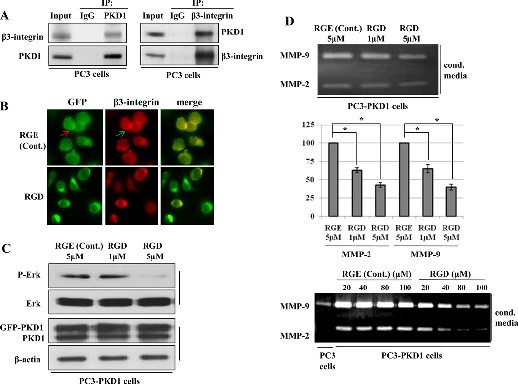
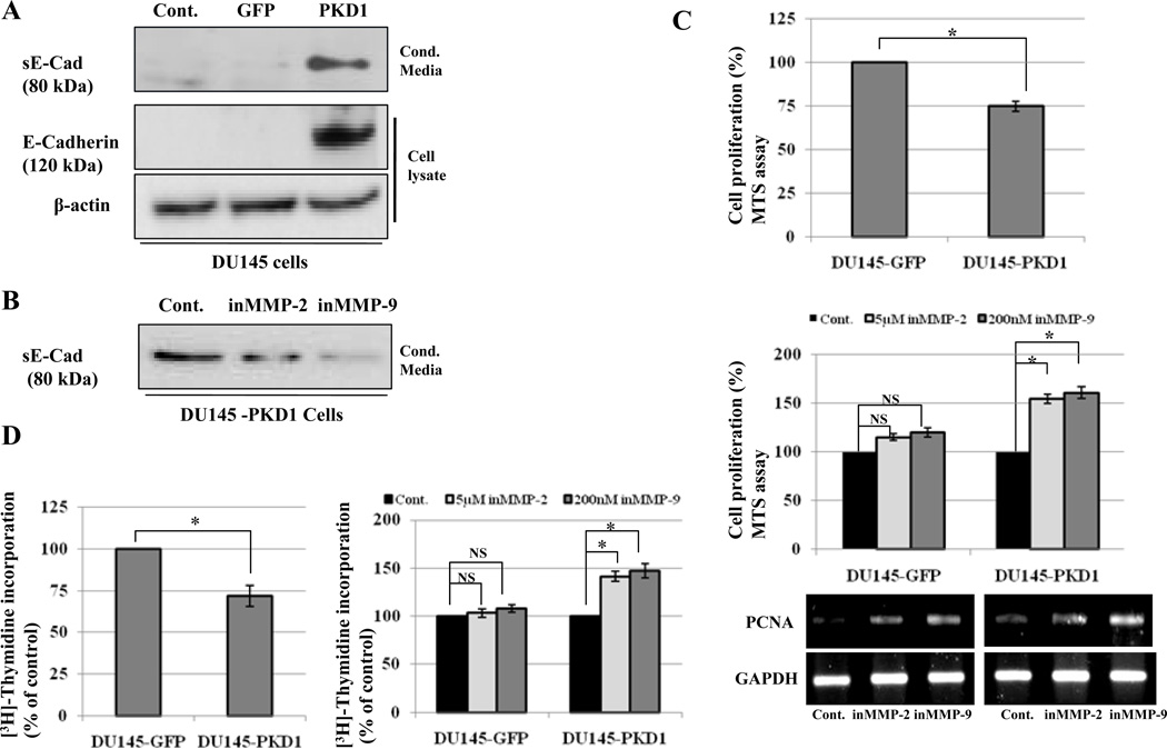
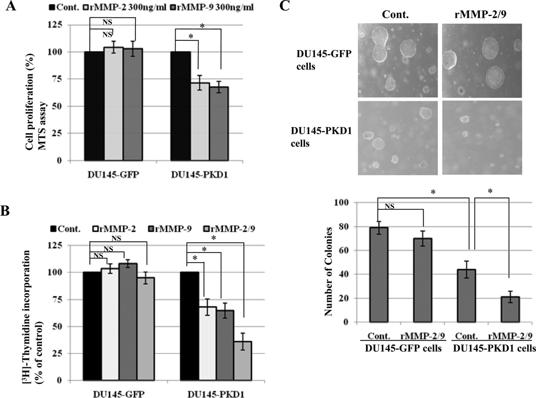
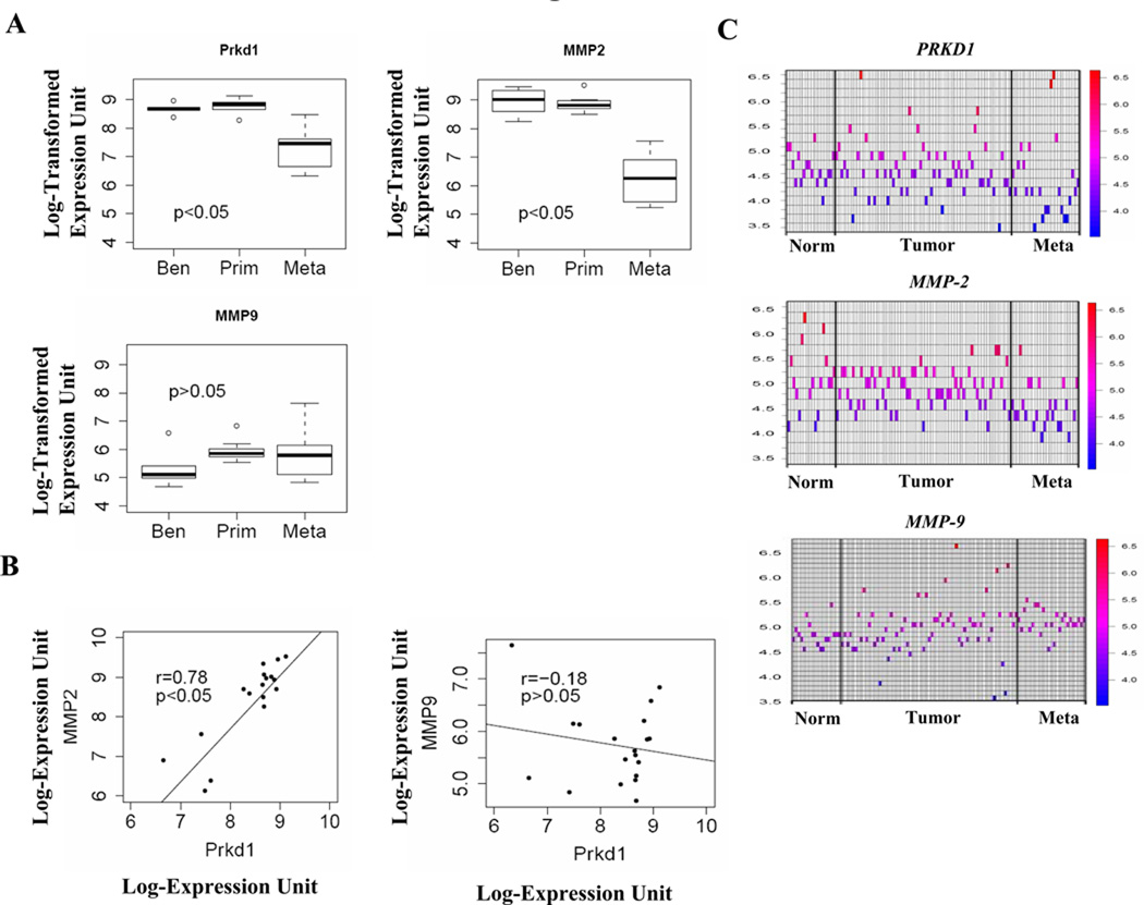
We and others previously showed that protein kinase D1 (PKD1) is downregulated in several cancers including prostate; interacts with E-cadherin, a major cell adhesion epithelial protein; and causes increased cell aggregation and decreased motility of prostate cancer cells. In this study, we show that PKD1 complexes with beta3-integrin, resulting in activation of mitogen-activated protein kinase/extracellular signal-regulated kinase (ERK) kinase-ERK pathway, which causes increased production of matrix metalloproteinase (MMP)-2 and MMP-9, that is associated with shedding of soluble 80 kDa E-cadherin extracellular domain. Interestingly, decreased cell proliferation following PKD1 transfection was rescued by MMP-2 and MMP-9 inhibitors and augmented by recombinant MMP-2 (rMMP-2) and rMMP-9 proteins, suggesting an antiproliferative role for MMPs in prostate cancer. Translational studies by in silico analysis of publicly available DNA microarray data sets show a significant direct correlation between PKD1 and MMP-2 expression in human prostate tissues. The study shows a novel mechanism for antiproliferative effects of PKD1, a protein of emerging translational interest in several human cancers, through increased production of MMP-2 and MMP-9 in cancer cells.
Figures
Similar articles
-
E-cadherin phosphorylation by protein kinase D1/protein kinase C{mu} is associated with altered cellular aggregation and motility in prostate cancer.Cancer Res. 2005 Jan 15;65(2):483-92. Cancer Res. 2005. PMID: 15695390
-
ADAM17 targets MMP-2 and MMP-9 via EGFR-MEK-ERK pathway activation to promote prostate cancer cell invasion.Int J Oncol. 2012 May;40(5):1714-24. doi: 10.3892/ijo.2011.1320. Epub 2011 Dec 23. Int J Oncol. 2012. PMID: 22200661
-
Shikonin inhibits prostate cancer cells metastasis by reducing matrix metalloproteinase-2/-9 expression via AKT/mTOR and ROS/ERK1/2 pathways.Int Immunopharmacol. 2014 Aug;21(2):447-55. doi: 10.1016/j.intimp.2014.05.026. Epub 2014 Jun 3. Int Immunopharmacol. 2014. PMID: 24905636
-
Emerging roles of protein kinase D1 in cancer.Mol Cancer Res. 2011 Aug;9(8):985-96. doi: 10.1158/1541-7786.MCR-10-0365. Epub 2011 Jun 16. Mol Cancer Res. 2011. PMID: 21680539 Free PMC article. Review.
-
Regulation of protein kinase D1 activity.Mol Pharmacol. 2012 Mar;81(3):284-91. doi: 10.1124/mol.111.075986. Epub 2011 Dec 21. Mol Pharmacol. 2012. PMID: 22188925 Free PMC article. Review.
Cited by
-
New pyrazolopyrimidine inhibitors of protein kinase d as potent anticancer agents for prostate cancer cells.PLoS One. 2013 Sep 23;8(9):e75601. doi: 10.1371/journal.pone.0075601. eCollection 2013. PLoS One. 2013. PMID: 24086585 Free PMC article.
-
Soluble E-cadherin: more than a symptom of disease.Front Biosci (Landmark Ed). 2012 Jan 1;17(5):1948-64. doi: 10.2741/4031. Front Biosci (Landmark Ed). 2012. PMID: 22201848 Free PMC article. Review.
-
Novel In Vivo model for combinatorial fluorescence labeling in mouse prostate.Prostate. 2015 Jun 15;75(9):988-1000. doi: 10.1002/pros.22984. Epub 2015 Mar 8. Prostate. 2015. PMID: 25753731 Free PMC article.
-
Inducible silencing of protein kinase D3 inhibits secretion of tumor-promoting factors in prostate cancer.Mol Cancer Ther. 2012 Jul;11(7):1389-99. doi: 10.1158/1535-7163.MCT-11-0887. Epub 2012 Apr 24. Mol Cancer Ther. 2012. PMID: 22532599 Free PMC article.
-
Potential role for protein kinase D inhibitors in prostate cancer.J Mol Med (Berl). 2023 Apr;101(4):341-349. doi: 10.1007/s00109-023-02298-4. Epub 2023 Feb 27. J Mol Med (Berl). 2023. PMID: 36843036 Review.
References
-
- Frisch SM, Ruoslahti E. Integrins and anoikis. Curr Opin Cell Biol. 1997;9:701–706. - PubMed
-
- Kantak SS, Kramer RH. E-cadherin regulates anchorage-independent growth and survival in oral squamous cell carcinoma cells. J Biol Chem. 1998;273:16953–16961. - PubMed
-
- Stetler-Stevenson WG, Krutzsch HC, Wacher MP, Margulies IM, Liotta LA. The activation of human type IV collagenase proenzyme. Sequence identification of the major conversion product following organomercurial activation. J Biol Chem. 1989;264:1353–1356. - PubMed
-
- McCawley LJ, Matrisian LM. Matrix metalloproteinases: they're not just for matrix anymore! Curr Opin Cell Biol. 2001;13:534–540. - PubMed
Publication types
MeSH terms
Substances
Grants and funding
LinkOut - more resources
Full Text Sources
Medical
Miscellaneous
