. 2010 Mar 1;70(5):1885-95.
doi: 10.1158/0008-5472.CAN-09-2833. Epub 2010 Feb 16.

Talin1 promotes tumor invasion and metastasis via focal adhesion signaling and anoikis resistance

Affiliations

Talin1 promotes tumor invasion and metastasis via focal adhesion signaling and anoikis resistance

Shinichi Sakamoto et al. Cancer Res. .

Abstract

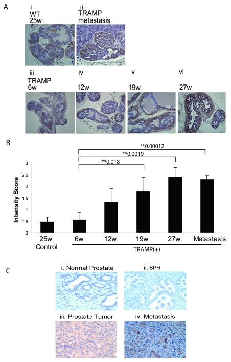
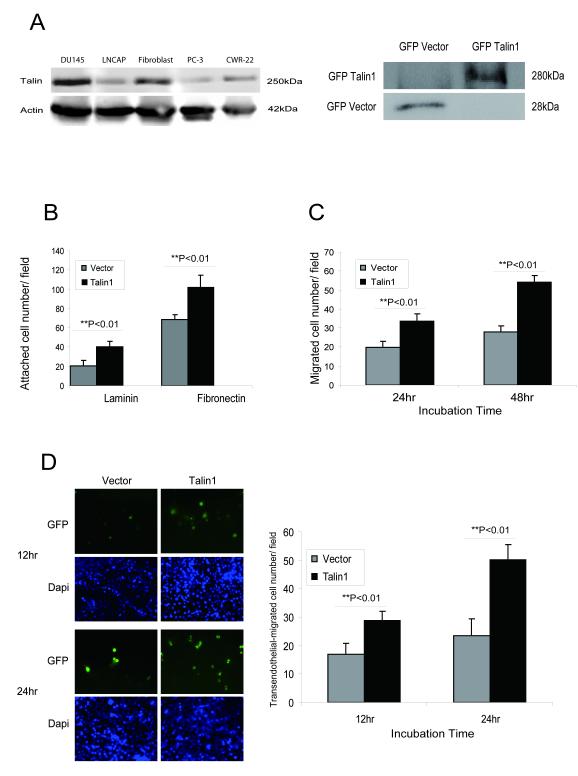
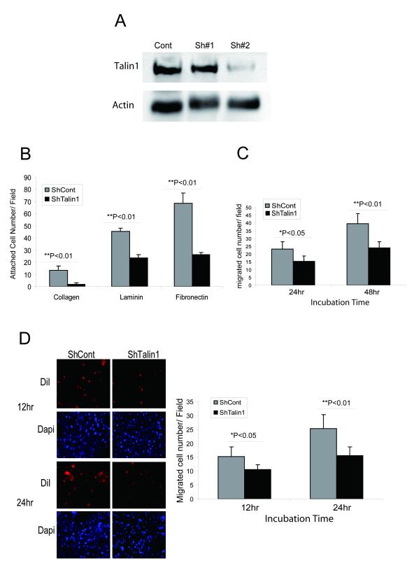
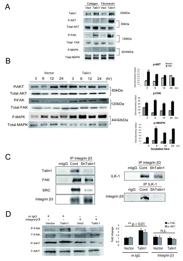
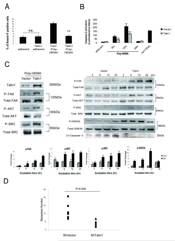
Talin1 is a focal adhesion complex protein that regulates integrin interactions with ECM. This study investigated the significance of talin1 in prostate cancer progression to metastasis in vitro and in vivo. Talin1 overexpression enhanced prostate cancer cell adhesion, migration, and invasion by activating survival signals and conferring resistance to anoikis. ShRNA-mediated talin1 loss led to a significant suppression of prostate cancer cell migration and transendothelial invasion in vitro and a significant inhibition of prostate cancer metastasis in vivo. Talin1-regulated cell survival signals via phosphorylation of focal adhesion complex proteins, such as focal adhesion kinase and Src, and downstream activation of AKT. Targeting AKT activation led to a significant reduction of talin1-mediated prostate cancer cell invasion. Furthermore, talin1 immunoreactivity directly correlated with prostate tumor progression to metastasis in the transgenic adenocarcinoma mouse prostate mouse model. Talin1 profiling in human prostate specimens revealed a significantly higher expression of cytoplasmic talin1 in metastatic tissue compared with primary prostate tumors (P < 0.0001). These findings suggest (a) a therapeutic significance of disrupting talin1 signaling/focal adhesion interactions in targeting metastatic prostate cancer and (b) a potential value for talin1 as a marker of tumor progression to metastasis.


Figures

Figure 1
Figure 1. Talin1 Profiling during Prostate Tumor Progression in the TRAMP Model and human Prostate Cancer Specimens
Panel (A), Talin1 expression was analyzed in paraffin-embedded prostate tissue specimens from TRAMP mice and control age-matched mice (6-27 wks). A strong immunoreactivity for talin1 was detected in prostate tumors derived from both 19wk and 27wk TRAMP mice, subpanels (iv) and (v), respectively. Sub-panel (ii) represents a metastatic lesion to the liver in a TRAMP mouse (27wks). Panel (B), The intensity of IHC staining (H) was calculated by P=Percentage of staining × I intensity (1-3). The numerical data reveal a positive correlation between increasing tumor grade and progression to metastasis and increased talin expression. (*) and (**) indicate statistical difference at p value<0.05 and <0.01, respectively, compared to specimens from control mice. Panel (C); Talin1 immunoreactivity was analyzed in paraffin-embedded prostate tissue specimens from normal (i), BPH (ii), primary prostate tumors (iii) and metastasis (iv).
Figure 2
Figure 2. Talin1 Enhances Cell Adhesion, Migration and Invasion
Panel A, Talin1 endogenous expression in human prostate cancer cell lines; actin expression is used as a loading control. Bands detected indicate GFP-talin1 (280kDa) and GFP-vector protein (28kDa) respectively. Panel B, Cell adhesion potential was comparatively analyzed in laminin or fibronectin-coated plates in PC-3 GFP vector control and PC-3 GFP- talin1 cloned transfectants. Values represent the mean from three indepeden experiments performed duplicate ± SEM. Panel C, PC-3 cells were subjected to wounding and migrating cells were counted under the microscope in three independent fields (3 wells/condition). Panel D, The transendothelial migration assay was performed using HBMECs, and either PC-3 GFP-vector or PC-3 GFP-talin1 expressing cells, as described in “Materials and Methods”. Green and blue staining indicated GFP vector/talin1 cells and DAPI nuclear staining, respectively. Numerical data represent values from three independent experiments ± SEM.
Figure 3
Figure 3. Talin1 Loss Decreases Cell Adhesion, Migration and Invasion
Panel A, ShRNA talin1 stable cell line was established in DU-145 cells. Panel B, ShRNA vector and ShRNA-talin1 transfectant cells were comparatively analyzed for their adhesion potential (as described above). Values represent mean ± SEM from three independent experiments. Panel C, Talin1 silencing reduces DU-145 cell migration ability. Cells were subjected to wounding and their migration potential was evaluated; the number of migrating cells was counted under the microscope in three independent fields (40×***) per well (3 wells/condition). Panel D, Loss of talin1 inhibits prostate tumor cell invasion. Transendothelial migration assay was conducted using HBMEC (human brain microvascular endothelial cell) DU-145 vector and ShRNA Talin1 stable cell line. The number of invading cells (red) was determined under confocal microscopy after 12 and 24 hrs. Blue color represents DAPI nuclear staining.
Figure 4
Figure 4. Talin1 Mediates ECM-dependent Activation of FAK/AKT Pathway
Panel A, After adherence of GFP-talin1 and vector-transfected PC-3 cells on either collagen or fibronectin plates (6hrs), cells were lysed and subjected to Western blot analysis. Levels of total and phosphorylated AKT (Ser473), FAK (Y397) and MAPK proteins were detected using respective antibodies. Panel B, Phosphorylation of AKT (Ser473), FAK(Y397) and MAPK were evaluated after (6hr) adherence to fibronectin. Quantification of phosphorylation was performed by densitometry (as shown on the right). Panel C, ShTalin1 DU-145 cells were subjected to immunoprecipitation using the integrin β3 antibody or the ILK-1 antibody. As controls, proteins were precipitated with the control IgG derived from the same species plus the secondary antibody (protein G/A). Talin1, total FAK and SRC binding were detected by Western blotting. Binding of ILK-1 or integrin β3 was determined using the respective antibodies (right panel). Panel D, GFP vector and talin1 expressing PC-3 cells were plated on fibronectin-coated plate incubated with mouse IgG or integrin β3 (blocking) antibodies (20μg/ml); The total proteins and phosphorylated forms of FAK and AKT were determined by immunblotting after 6hrs of plating.
Figure 5
Figure 5. Talin1 Mediates Anoikis Resistance
Panel A, GFP-talin1 and vector PC-3 cells were kept in suspension conditions using Poly-HEMA coated plates for 24hr and subjected to Flow Cytometry using AnnexinV antibody. Panel B, Caspase-3 activation was measured in Poly-HEMA suspension conditions after 6, 12 and 24hrs as described in “Materials and Methods”. GFP talin1 and GFP vector cells were also treated with TRAIL (100ng/ml) plus Velcade (100nM) (positive control). Values indicate the mean from two independent experiments performed in triplicate ± SEM. (**) indicates statistically significant difference at p<0.01 Panel C, GFP-talin1 and vector PC-3 cells were kept in suspension conditions and after 24hrs, cells were lysed and subjected to Western blotting. Expression of total protein and phosphorylated forms of AKT (Ser473), FAK (Y397) and p42/44MAPK were detected using respective antibodies (left panel). Levels of total and phosphorylated AKT (Ser473), FAK (Y397), SRC and GSK3β as well as cleaved caspase-3, were assessed during different time points (3-24hr) (right panel). The phosphorylation status of individual proteins was evaluated by densitometry (lower panel). Panel D, Experimental metastasis assay. Nude mice were injected intravenously in the tail vein with prostate cancer cells, DU-145 ShVector, (n=6) and DU-145 shRNA talin1, [n=5, (2×10⁶/mouse)]. At 5-wks post-inoculation, lungs were dissected and metastastic lesions were counted. Talin 1 loss led to a significantly reduced number of metastatic lesions to the lungs (P =0.028).

Similar articles

Cited by

References

    1. Fornaro M, Manes T, Languino LR. Integrins and prostate cancer metastases. Cancer Metastasis Rev. 2001;20(34):321–31. - PubMed
    1. McKenzie S, Kyprianou N. Apoptosis evasion: the role of survival pathways in prostate cancer progression and therapeutic resistance. J Cell Biochem. 2006;97(1):18–32. - PMC - PubMed
    1. Frisch SM, Screaton RA. Anoikis mechanisms. Current opinion in cell biology. 2001;13(5):555–62. - PubMed
    1. Rennebeck G, Martelli M, Kyprianou N. Anoikis and survival connections in the tumor microenvironment: is there a role in prostate cancer metastasis? Cancer research. 2005;65(24):11230–5. - PMC - PubMed
    1. Giancotti FG, Ruoslahti E. Integrin signaling. Science. 1999;285(5430):1028–32. - PubMed

Publication types

MeSH terms