. 2010 Mar 2;107(9):4293-8.
doi: 10.1073/pnas.0905877107. Epub 2010 Feb 16.

TGF-beta regulates T-cell neurokinin-1 receptor internalization and function

Affiliations

TGF-beta regulates T-cell neurokinin-1 receptor internalization and function

Martin Beinborn et al. Proc Natl Acad Sci U S A. .

Abstract

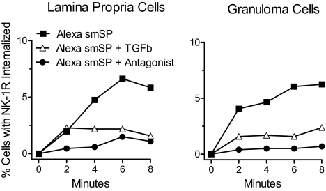
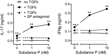
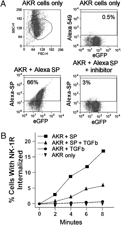
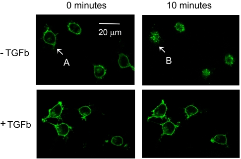
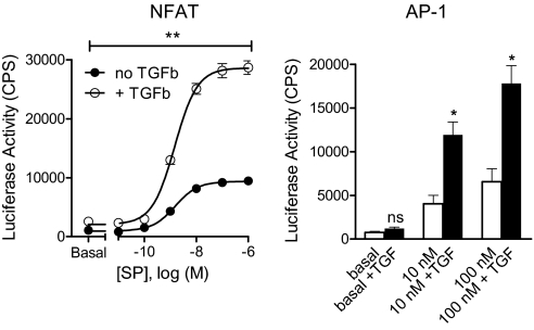
Substance P (SP) is a proinflammatory mediator implicated in inflammatory bowel disease (IBD) and other inflammatory states. SP acts by stimulating the neurokinin-1 receptor (NK-1R) on T lymphocytes and other cell types, and regulates these cells in a complex interplay with multiple cytokines. The mechanisms of interaction among these inflammatory mediators are not yet fully understood. Here, we demonstrate that function of the NK-1R, a member of the G protein-coupled receptor (GPCR) superfamily, is modulated by TGF-beta. The latter acts not on a GPCR but via serine-threonine kinase-class receptors. By flow confocal image analysis, we demonstrate that TGF-beta delays SP-induced NK-1R internalization on mucosal T cells isolated from a mouse model of IBD and on granuloma T cells in murine schistosomiasis. Furthermore, luciferase reporter-gene assays revealed that NK-1R stimulation activates the nuclear factor of activated T cell- and activator protein-1-dependent signaling pathways, which are known triggers of effector T-cell cytokine production. TGF-beta markedly increases SP-induced activation of these signaling cascades, suggesting that delayed NK-1R internalization results in enhanced signaling. Providing a link to amplified immune function, SP and TGF-beta, when applied in combination, trigger a strong release of the proinflammatory cytokines IFN-gamma and IL17 from intestinal inflammatory T cells, whereas either agonist alone shows no effect. These observations establish precedent that members of two distinct receptor superfamilies can interact via a previously unrecognized mechanism, and reveal a paradigm of GPCR transregulation that is relevant to IBD and possibly other disease processes.


Conflict of interest statement

The authors declare no conflict of interest.

Figures

Fig. 1.
Fig. 1.
Lamina propria T cells from colitic mice and granuloma T cells from mice infected with S. mansoni display slow NK-1R internalization following TGF-β (TGFb) exposure. T cells were isolated from the intestinal lamina propria of IL10−/− mice with colitis (Left) or from granulomas isolated from the liver of mice infected for 8 weeks with S. mansoni (Right). The T cells were preincubated with TGF-β or cultured alone for 18 h before exposure to the Alexa-labeled SP analog smSP (Alexa smSP, 10−9 M). This peptide binds only the NK1 tachykinin receptor with high-affinity. ImageStream analysis shows that TGF-β pre-exposure decreases smSP-induced NK-1R internalization. The smSP receptor-induced internalization was blocked when cells were cocultured with a highly specific NK-1R receptor antagonist (SR140333, 10−8 M). Data are representative results from multiple determinations. TGF-β effects on agonist-induced NK-1R internalization at early time points in several independent experiments are statistically compared in Fig. S1.
Fig. 2.
Fig. 2.
After TGF-β (TGFb) exposure, SP induces LPMC to secrete IL17 and IFN-γ. LPMC isolated from IL10−/− mice were cultured 24 h with or without TGF-β (5 ng/mL). The cells were washed and then placed into fresh medium with or without SP, in the presence or absence of NK-1R antagonist (SR140333, 10−8 M). After another 24 h, IL17 and IFN-γ in the culture medium were measured by ELISAs. Data are the mean ± SEM of quadruplicate determinations, and are representative of three independent experiments. **, P < 0.01 vs. secretion in the absence of TGF-β.
Fig. 3.
Fig. 3.
TGF-β (TGFb) delays NK-1R internalization following SP engagement. AKR T cells were stably transfected to express an NK-1R-eGFP fusion protein, and were analyzed by either flow cytometry (A) or ImageStream analysis (B). (A) Flow cytometry shows that AKR cells express eGFP (Right Upper) and that Alexa-labeled SP (Alexa) bound to the majority of these cells (Left Lower). This binding was specific for NK-1R engagement, because a highly specific NK-1R inhibitor (SR140333) blocked Alexa-SP binding (Right Lower). (B) ImageStream analysis reveals that exposing AKR cells to SP at 10−9 M induces NK1R-eGFP internalization. This is reflected by a time-dependent increase in the percentage of cells with fluorescent SP/NK-1R clusters (% cells with NK-1R internalized). Cells preincubated for 18 h with TGF-β show much slower internalization of NK-1R following SP engagement. AKR T cells with or without prior TGF--β exposure and no SP contact display little, if any, NK-1R-eGFP internalization over the observation period. TGF-β effects on agonist-induced NK-1R internalization at early time points in several independent experiments are statistically compared in Fig. S1.
Fig. 4.
Fig. 4.
TGF-β (TGFb) markedly delays agonist-induced NK-1R internalization. HEK293 cells stably expressing NK-1R-eGFP were incubated overnight on microscopy coverslips, either in the absence or presence of 5 ng/mL TGF-β. After mounting to the confocal microscopy stage, SP (10 nM) was added at time “0 minutes,” and fluorescent images were taken every 30 s. Within 10 min, SP induces receptor internalization (Upper pair of panels), which is in large part prevented by TGF-β (Lower pair of panels). One cell that is highlighted by arrows (“A” before and “B” after SP exposure) is shown at higher magnification in Fig. S4).
Fig. 5.
Fig. 5.
TGF-β (TGFb) enhances NK-1R-induced activation of NFAT and AP-1 promoters. HEK293 cells were transiently transfected with cDNAs encoding (i) NK-1R-eGFP, (ii) either NFAT-luciferase (Left) or AP-1-luciferase (Right), and (iii) a lac-Z control gene for data normalization. The cells were grown overnight either in the absence or presence of 5 ng/mL TGF-β, and were then washed and stimulated for 4 h with the indicated concentrations of SP. Data represent the mean ± SEM of three experiments. *, P < 0.05; **, P < 0.01; comparison of SP-induced activity in the absence vs. presence of TGF-β.

References

    1. O'Connor TM, et al. The role of substance P in inflammatory disease. J Cell Physiol. 2004;201:167–180. - PubMed
    1. Almeida TA, et al. Tachykinins and tachykinin receptors: structure and activity relationships. Curr Med Chem. 2004;11:2045–2081. - PubMed
    1. Smith VC, Sagot MA, Couraud JY, Buchan AM. Localization of the neurokinin 1 (NK-1) receptor in the human antrum and duodenum. Neurosci Lett. 1998;253:49–52. - PubMed
    1. Goode T, et al. Substance P (neurokinin-1) receptor is a marker of human mucosal but not peripheral mononuclear cells: molecular quantitation and localization. J Immunol. 1998;161:2232–2240. - PubMed
    1. Cook GA, et al. Molecular evidence that granuloma T lymphocytes in murine schistosomiasis mansoni express an authentic substance P (NK-1) receptor. J Immunol. 1994;152:1830–1835. - PubMed

Publication types