. 2010 Mar 2;107(9):4305-10.
doi: 10.1073/pnas.0909024107. Epub 2010 Feb 16.

Islet amyloid deposition limits the viability of human islet grafts but not porcine islet grafts

Affiliations

Islet amyloid deposition limits the viability of human islet grafts but not porcine islet grafts

K J Potter et al. Proc Natl Acad Sci U S A. .

Abstract

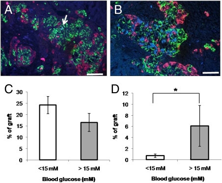
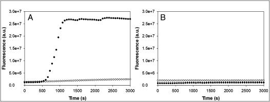
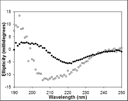
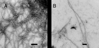
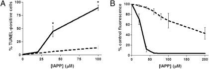
Islet transplantation is a promising treatment for diabetes but long-term success is limited by progressive graft loss. Aggregates of the beta cell peptide islet amyloid polypeptide (IAPP) promote beta cell apoptosis and rapid amyloid formation occurs in transplanted islets. Porcine islets are an attractive alternative islet source as they demonstrate long-term graft survival. We compared the capacity of transplanted human and porcine islets to form amyloid as an explanation for differences in graft survival. Human islets were transplanted into streptozotocin-diabetic immune-deficient mice. Amyloid deposition was detectable at 4 weeks posttransplantation and was associated with islet graft failure. More extensive amyloid deposition was observed after 8 weeks. By contrast, no amyloid was detected in transplanted neonatal or adult porcine islets that had maintained normoglycemia for up to 195 days. To determine whether differences in IAPP sequence between humans and pigs could explain differences in amyloid formation and transplant viability, we sequenced porcine IAPP. Porcine IAPP differs from the human sequence at 10 positions and includes substitutions predicted to reduce its amyloidogenicity. Synthetic porcine IAPP was considerably less amyloidogenic than human IAPP as determined by transmission electron microscopy, circular dichroism, and thioflavin T binding. Viability assays indicated that porcine IAPP is significantly less toxic to INS-1 beta cells than human IAPP. Our findings demonstrate that species differences in IAPP sequence can explain the lack of amyloid formation and improved survival of transplanted porcine islets. These data highlight the potential of porcine islet transplantation as a therapeutic approach for human diabetes.


Conflict of interest statement

The authors declare no conflict of interest.

Figures

Fig. 1.
Fig. 1.
Rapid amyloid formation is associated with human islet graft failure. Human islets were grafted in streptozotocin-diabetic NOD/SCID recipients as described in Materials and Methods (n = 43). Small amounts of amyloid (arrow) were detected by thioflavin S stain (blue) in grafts in normoglycemic recipients at 4 weeks posttransplant (A) but were more marked at 8 weeks posttransplant and in hyperglycemic recipients (B). Amyloid appeared adjacent to insulin-positive cells (green) and areas of apparent islet cell loss, but not glucagon-positive cells (red). (Scale bar, 50 μm.) Beta cell area (C) tended to be reduced and amyloid area was increased (D) in recipients of grafts with blood glucose values >15 mM at the time of graft harvest. The number of recipients in the normoglycemic and hyperglycemic recipients were 31 and 12, respectively. *, denotes statistically significant difference from normoglycemic (<15 mM) group (P < 0.05).
Fig. 2.
Fig. 2.
Lack of amyloid in native or transplanted porcine islets. (A) Human islet. (B) Human islet graft. (C) Adult porcine islet. (D) Neonatal porcine islet graft. (E) Adult porcine islet graft. Amyloid is detectable by thioflavin S (blue) stain in human but not adult porcine islets. Amyloid develops rapidly in transplanted human islets but is not found in transplanted neonatal or adult porcine islets. Red immunostaining, insulin. (Scale bar, 50 μm.)
Fig. 3.
Fig. 3.
Primary sequences of human and porcine islet amyloid polypeptide. (A) Human IAPP (hIAPP) sequence. (B) Porcine IAPP (pIAPP) sequence. All peptides have an amidated C terminus and free N terminus with a Cys-2 to Cys-7 disulfide bridge. Residues are numbered according to their position in mature IAPP. Residues differing from the human sequence are indicated in bold.
Fig. 4.
Fig. 4.
Kinetics of human and porcine IAPP aggregation. (A) Thioflavin-T monitored aggregation kinetics of human (black circles) and porcine (white circles) IAPP indicate that porcine IAPP is less amyloidogenic than its human counterpart. (B) Thioflavin-T kinetics of porcine IAPP seeded by human IAPP aggregates. Seeding experiments were conducted by adding porcine IAPP to a solution containing either 3.2 μM (black circles) or 6.4 μM (white circles) preformed human IAPP amyloid fibrils. All reactions were monitored at pH 7.4 in 2% HFIP and 25 °C. Peptide concentration was 32 μM. Experiments were conducted with constant stirring to maintain solution homogeneity. Experiments were performed a minimum of three separate times and similar results were obtained.
Fig. 5.
Fig. 5.
Far UV CD spectra of human and porcine IAPP. Conformational analysis of human (black circles) and porcine (white circles) IAPP after 12-h incubation indicates that porcine IAPP has significantly less β-sheet propensity than the human peptide. Human IAPP adopts a classic β-sheet conformation signified by a signal minima at 218 nm and positive ellipticity below 200 nm. In contrast, porcine IAPP shows a wide band ranging from 208 to 222 nm, indicative of a mixture of helical and β-sheet structures. All reactions were monitored at pH 7.4 in 2% HFIP and 25 °C . Peptide concentration was 32 μM. Spectra are the average of three repeats. All samples contained 16 mM Tris HCl.
Fig. 6.
Fig. 6.
TEM micrographs of human and porcine IAPP at physiological pH. (A) After 24 h of incubation, human IAPP samples appear as dense mats of intertwined amyloid fibrils with the classic linear and unbranched morphology. (B) Porcine IAPP samples incubated for the same length of time show the presence of a few fibrillar strands among a bed of amorphous aggregates. TEM samples were incubated at pH 7.4 in 2% HFIP and 25 °C. Peptide concentration was 32 μM. (Scale bar, 100 nm.)
Fig. 7.
Fig. 7.
Porcine IAPP is less cytotoxic than human IAPP. (A) TUNEL staining following 16-h incubation of INS-1 cells in the presence of human or porcine IAPP. *, denotes statistically significant difference from porcine IAPP-treated cells (P < 0.05). (B) Alamar blue viability assay following 24-h incubation of INS-1 beta cells in the presence of human or porcine IAPP. The EC50 for human IAPP was 27 μM compared to 172 μM for porcine IAPP (P < 0.05). Solid line, human IAPP; dashed line, porcine IAPP.

References

    1. Shapiro AM, et al. International trial of the Edmonton protocol for islet transplantation. N Engl J Med. 2006;355:1318–1330. - PubMed
    1. Menger MD, Yamauchi J, Vollmar B. Revascularization and microcirculation of freely grafted islets of Langerhans. World J Surg. 2001;25:509–515. - PubMed
    1. Avila JG, et al. Improved outcomes in islet isolation and transplantation by the use of a novel hemoglobin-based O2 carrier. Am J Transplant. 2006;6:2861–2870. - PubMed
    1. Lee Y, et al. Metabolic mechanisms of failure of intraportally transplanted pancreatic beta-cells in rats: Role of lipotoxicity and prevention by leptin. Diabetes. 2007;56:2295–2301. - PubMed
    1. Noguchi H, et al. Activation of c-Jun NH2-terminal kinase (JNK) pathway during islet transplantation and prevention of islet graft loss by intraportal injection of JNK inhibitor. Diabetologia. 2007;50:612–619. - PubMed

Publication types