. 2010 Mar 2;107(9):4412-7.
doi: 10.1073/pnas.1000738107. Epub 2010 Feb 16.

Endocytosis and clathrin-uncoating defects at synapses of auxilin knockout mice

Affiliations

Endocytosis and clathrin-uncoating defects at synapses of auxilin knockout mice

Yang-In Yim et al. Proc Natl Acad Sci U S A. .

Abstract

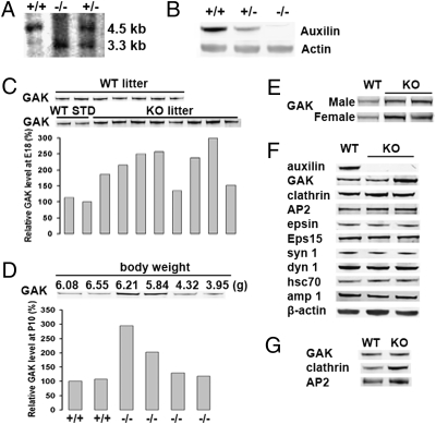
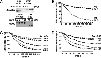
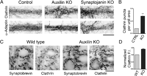
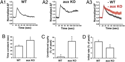
Neuronally expressed auxilin and ubiquitously expressed cyclin-G-dependent kinase (GAK) are homologous proteins that act as cochaperones to support the Hsc70-dependent clathrin uncoating of clathrin-coated vesicles. GAK was previously shown to be essential in mouse during embryonic development and in the adult. We have now engineered an auxilin knockout mouse. Mutant mice had a high rate of early postnatal mortality and surviving pups generally had a lower body weight than wild-type pups, although they had a normal life span. GAK was up-regulated as much as 3-fold in the brains of both surviving neonates and adult mutant mice. An increased number of clathrin-coated vesicles and empty cages were present at knockout synapses both in situ and in primary neuronal cultures. Additionally, clathrin-mediated endocytosis of synaptic vesicles in knockout hippocampal neurons was impaired, most likely due to sequestration of coat components in assembled coats and cages. Collectively, our results demonstrate the specialized role of auxilin in the recycling of synaptic vesicles at synapses, but also show that its function can be partially compensated for by up-regulation of GAK.


Conflict of interest statement

The authors declare no conflict of interest.

Figures

Fig. 1.
Fig. 1.
Analysis of endocytic proteins in auxilin knockout mice. (A) Southern blot analysis of knockout mice. The 5′ probe detects a 3.3-kb and a 4.5-kb band for the mutant and wild-type alleles, respectively. (B) Western blot analysis of auxilin from auxilin knockout mice. The brain cytosols from each genotype were analyzed to check the auxilin level. (C) GAK expression level in brain lysates of embryonic wild-type and auxilin knockout mice (E18). Brain lysates from embryos of a single wild-type (WT) litter and a single knockout (KO) litter, obtained from first-time mothers, were run on Western blots and then immunoblotted for GAK. In the western blot of GAK levels in the KO litter, the first two lanes are WT brain lysate standards (STD). The GAK intensity in KO brain lysates was plotted in the bar graph as relative GAK level by normalizing the intensity of the GAK bands to that of the WT lysate in lane 2. The same amount of protein from brain lysates was loaded in each lane. (D) Relationship between body weight and GAK expression level in auxilin knockout pups. Brain lysates from a single KO litter (P10) were analyzed for GAK level by Western blot. For comparison, brain lysates from a WT litter (P10) were run in lanes 1 and 2. The weights of the auxilin KO mice are shown above the blot, whereas the genotypes are shown below the graph. The relative GAK expression level is plotted directly beneath the immunoblot. (E) Western blots of GAK in neuronal tissue of wild-type and auxilin knockout mice at 5 weeks of age. (F) Western blots of the brain extract from wild-type and auxilin knockout mice at P7. Clathrin-mediated endocytosis-related proteins were detected from each brain extract using indicated antibodies. (G) Increased expression of clathrin and AP2 in neuronal tissue of auxilin knockout runts. Western blot for AP2 and clathrin of brain cytosol was obtained from a wild-type pup (P7) and an auxilin knockout runt (P7).
Fig. 2.
Fig. 2.
Effect of knocking out auxilin on uncoating activity of brain cytosol. (A) Expression level of auxilin and GAK in wild-type brain cytosol from mice at P0.5, P3, P7, and P21. Western blots were used to determine the auxilin and GAK levels in brain cytosol by using protein standards (10 and 50 nM) of auxilin (Aux-C54) and GAK (GAK-C62). The auxilin and GAK were immunoblotted using anti-auxilin and anti-GAK antibodies, respectively. The same amount of protein from brain lysates was loaded in each lane. (B) Kinetics of clathrin uncoating by brain cytosol from wild-type and auxilin knockout mice. Clathrin-uncoating assays were conducted as described inMethods using permeabilized cells expressing GFP-clathrin. Brain cytosols (1 mg/200 μL) from newborn wild-type mice (P0.5) and auxilin knockout mice (P0.5) were used in the assay. (C) Time course of clathrin uncoating by Hsc70 as a function of Aux-C54 concentration. Varying concentrations of Aux-C54 (5–100 nM) were added to 2 μM Hsc70 and 1 mM ATP in the uncoating assay. (D) Time course of clathrin uncoating by Hsc70 as a function of GAK-C62 concentration. Varying concentrations of GAK-C62 (10–800 nM) were added to 2 μM Hsc70 and 1 mM ATP in the uncoating assay.
Fig. 3.
Fig. 3.
Immunoreactivity for endocytic clathrin coat components is clustered at auxilin knockout synapses, and clustering resembles the pattern observed in synaptojanin 1 knockout synapses. Immunofluorescence pictures are shown as negative images (fluorescence is shown in black). (A) High-magnification views of cultured neurons from an auxilin knockout mouse, a littermate pup, and a synaptojanin 1 knockout mouse immunostained for clathrin and AP2 (with α-adaptin antibodies). In the two knockout images, the clustering of clathrin and AP2 immunofluorescence, which reflects accumulations of clathrin-coated structures at synapses (16), is increased. (Scale bar, 20 μm.) (B) Number of clathrin puncta above threshold per unit area in auxilin knockout cultures. (C) Sections of deep cerebellar nuclei from auxilin knockout and wild-type mice immunostained for clathrin and for the synaptic vesicle marker synaptobrevin. Note that clathrin fluorescence, but not synaptobrevin fluorescence, is more intense at knockout synapses. (Scale bar, 20 μm.) (D) Clathrin immunofluorescence intensity (F.I.) normalized to the synaptobrevin immunofluorescence intensity. Error bars in B and C are SEM.
Fig. 4.
Fig. 4.
Accumulation of CCVs and clathrin cages at auxilin knockout synapses from deep cerebellar nuclei of adult mice as revealed by electron microscopy. (A) Representative example of a wild-type synapse. A clathrin-coated structure is indicated by the circle. (B) Percentage of CCVs/cages (assembled clathrin structures, CSs) relative to synaptic vesicles (SVs) at wild-type and knockout synapses. (C and D) Examples of auxilin KO synapses where a variable number of CCVs or empty cages can be seen. Typically, CCVs/cages (asterisks indicate areas enriched in these structures) are located at the periphery of synaptic vesicle clusters (such clusters are delineated by dotted lines in the figures) and often along the plasma membrane. The insets in C and D are high-magnification views of the rectangular regions that show empty cages and CCVs, respectively. (Scale bars, 200 nm.)
Fig. 5.
Fig. 5.
Auxilin knockout reduces the rate of endocytosis. (A) Representative examples of synaptopHluorin signal from a single hippocampal bouton (A1 and A2) and the averaged fluorescence trace (A3); 77 boutons from 4 wild-type mice and 48 boutons from 4 auxilin knockout mice after 200 AP at 20 Hz in WT (black) and auxilin KO (red) neurons. Traces are normalized to the basal fluorescence recorded before stimulation. The decay of the fluorescence signal indicates endocytosis. (B) The endocytic time constant of hippocampal boutons after 200 action potentials at 20 Hz in WT mice (n = 27) and auxilin KO mice (n = 14); *P = 0.01. (C) The percentage of fluorescence that remained unretrieved after 120 s in WT and auxilin KO mice; *P = 0.0045. The data were normalized to the peak of the fluorescence increase. (D) The initial endocytosis rate within 10 s after stimulation in WT and auxilin KO mice; *P = 0.0184. Error bars are SEM.

Similar articles

Cited by

References

    1. Ehrlich M, et al. Endocytosis by random initiation and stabilization of clathrin-coated pits. Cell. 2004;118:591–605. - PubMed
    1. Loerke D, et al. Cargo and dynamin regulate clathrin-coated pit maturation. PLoS Biol. 2009;7:e57. - PMC - PubMed
    1. Schlossman DM, Schmid SL, Braell WA, Rothman JE. An enzyme that removes clathrin coats: Purification of an uncoating ATPase. J Cell Biol. 1984;99:723–733. - PMC - PubMed
    1. Chappell TG, et al. Uncoating ATPase is a member of the 70 kilodalton family of stress proteins. Cell. 1986;45:3–13. - PubMed
    1. Sousa R, Lafer EM. Keep the traffic moving: Mechanism of the Hsp70 motor. Traffic. 2006;7:1596–1603. - PMC - PubMed

Publication types