. 2010 Mar 2;107(9):4317-22.
doi: 10.1073/pnas.0910261107. Epub 2010 Feb 16.

Surgery with molecular fluorescence imaging using activatable cell-penetrating peptides decreases residual cancer and improves survival

Affiliations

Surgery with molecular fluorescence imaging using activatable cell-penetrating peptides decreases residual cancer and improves survival

Quyen T Nguyen et al. Proc Natl Acad Sci U S A. .

Abstract

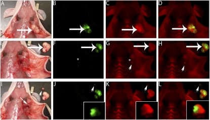
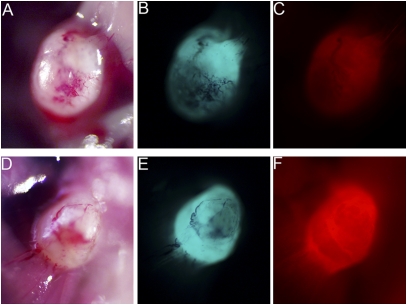
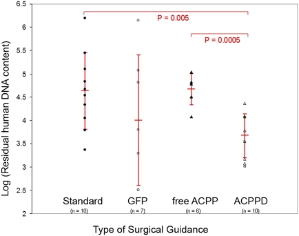
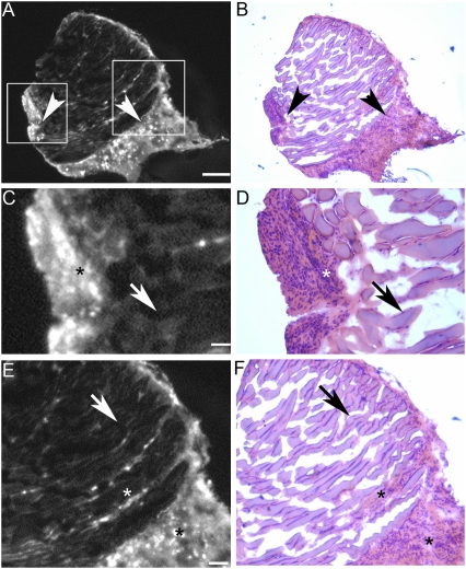
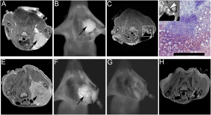
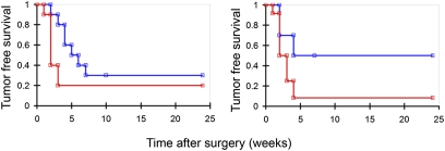
The completeness of tumor removal during surgery is dependent on the surgeon's ability to differentiate tumor from normal tissue using subjective criteria that are not easily quantifiable. A way to objectively assess tumor margins during surgery in patients would be of great value. We have developed a method to visualize tumors during surgery using activatable cell-penetrating peptides (ACPPs), in which the fluorescently labeled, polycationic cell-penetrating peptide (CPP) is coupled via a cleavable linker to a neutralizing peptide. Upon exposure to proteases characteristic of tumor tissue, the linker is cleaved, dissociating the inhibitory peptide and allowing the CPP to bind to and enter tumor cells. In mice, xenografts stably transfected with green fluorescent protein show colocalization with the Cy5-labeled ACPPs. In the same mouse models, Cy5-labeled free ACPPs and ACPPs conjugated to dendrimers (ACPPDs) delineate the margin between tumor and adjacent tissue, resulting in improved precision of tumor resection. Surgery guided by ACPPD resulted in fewer residual cancer cells left in the animal after surgery as measured by Alu PCR. A single injection of ACPPD dually labeled with Cy5 and gadolinium chelates enabled preoperative whole-body tumor detection by MRI, intraoperative guidance by real-time fluorescence, intraoperative histological analysis of margin status by fluorescence, and postoperative MRI tumor quantification. Animals whose tumors were resected with ACPPD guidance had better long-term tumor-free survival and overall survival than animals whose tumors were resected with traditional bright-field illumination only.


Conflict of interest statement

Conflict of interest statement: Q.T.N., E.S.O., T.A.A., T.J., and R.Y.T. have signed a scientific advisory agreement with a company founded to develop the technology described in this article.

Figures

Fig. 1.
Fig. 1.
ACPPs delineate tumor at the margin of resection. (A) White light image of a MDA-MB 435 xenograft following skin incision and tumor (large arrow) exposure. (B) Fluorescence image of GFP-labeled tumor cells from the same animal as in A. (C) Fluorescence image 6 h following i.v. administration of Cy5-labeled free ACPP showing increased uptake by the tumor (large arrow) compared to surrounding tissue. (D) Overlay fluorescence image showing colocalization of the Cy5 free ACPP with the GFP-labeled tumor. Following gross tumor excision by standard (unguided) technique, the tumor bed (*) seen with white light (E) appears to be free of tumor [the excised tumor (large arrow) has been laid next to the mouse]. (F) Fluorescence imaging of the GFP signal in the tumor bed appears to confirm complete tumor excision (*). However, imaging of the Cy5 signal (G and H) demonstrates a residual fluorescence signal (arrowhead) in surrounding remaining tissue (i.e., surgical margin). Using the Cy5 fluorescence to guide exploration (G and H), a small piece of residual tumor (arrowhead) is identified buried underneath the pectoralis muscle (I, small arrow). Interestingly, once the tumor has been dissected out from its buried position under the pectoralis muscle, the GFP signal (J, arrowhead) confirming the presence of tumor cells can be visualized along with the Cy5 free ACPP signal (K and L). (JL, insets) The excised tumor magnified and brightened 5×.
Fig. 2.
Fig. 2.
Comparing free ACPP and ACPPD for fluorescence imaging guidance. White light (A and D) and fluorescence images showing GFP-labeled MDA-MB 435 xenografts (B and E) from mice that were treated with Cy5-labeled free ACPP (C) and ACPPD (F). In tumors of comparable size, treatment with the free ACPP (C) or ACPPD (F) resulted in tumor-specific fluorescence uptake. There is a higher tumor-to-background fluorescence contrast for the ACPPD compared to free ACPPs and a higher absolute tumor fluorescence for ACPPDs when normalized to Cy5 fluorescence injected. Cy5 fluorescence images (C and F) were adjusted for total nanomoles of Cy5 injected per animal.
Fig. 3.
Fig. 3.
ACPPD guidance results in fewer residual tumor cells quantified by Alu PCR. Scatter plot showing residual human DNA content (i.e., tumor cells) for each type of surgery (standard surgery with no molecular guidance, GFP guidance, Cy5 free ACPP guidance, Cy5–ACPPD guidance). Vertical bar for each type of surgical condition shows the log of the mean and the standard deviation of human cell number that remained following surgery from the cohort of mice. There is significantly fewer residual human DNA content for surgery with ACPPD guidance compared to any other surgical conditions.
Fig. 4.
Fig. 4.
Histological analysis of surgical specimens derived from ACPPD-guided surgery. Photomicrographs showing a representative specimen from the B16F10 isograft model that was excised with fluorescently labeled ACPPD guidance. (A) Low-power Cy5 fluorescence showing positive ACPPD uptake (arrowheads). (B) The same section as in A stained with H&E, confirming the presence of malignant cells in regions that show increased fluorescence uptake (arrowheads). (C and E) Enlarged fluorescence images from the boxed areas in A, showing the demarcation between high (*) and low (arrows) fluorescence uptake. (D and F) Histological (H and E) analysis of C and E, showing that the areas of high fluorescence uptake correspond to malignant cells (*). (Scale bar in A and B: 0.5 mm; C and D: 0.1 mm; E and F: 0.25 mm.)
Fig. 5.
Fig. 5.
Dual-Labeled ACPPD. (AD) Example of HT1080 xenograft treated with ACPPD dually labeled with gadolinium and Cy5. Preoperative MR image of mouse showing contrast uptake in tumor (A, black arrow). Following skin incision and retraction, the tumor (black arrow) on the left chest wall was visible with Cy5 fluorescence (B). Following initial surgery, repeat MRI (C) showed a small area of tissue with increased gadolinium uptake (D inset, white arrowhead). This area of tissue was identified using fluorescence imaging at a second surgery. Histological analysis of this tissue confirmed the presence of cancer cells (D). (Scale bar: 100 μm). (EH) Example of MDA-MB 435 xenograft treated with ACPPD dually labeled with gadolinium and Cy5. Preoperative MR image of mouse showing contrast uptake in tumor (E, black arrow). Following skin incision and retraction, the tumor (black arrow) on the left chest wall was visible with Cy5 fluorescence (F). Tumor was resected using ACPPD–Cy5 imaging guidance until all visible fluorescence was completely removed (G). Repeat MR imaging following surgery showed complete removal of all tumor (H).
Fig. 6.
Fig. 6.
Kaplan–Meier survival curve. Tumor-free survival after surgery versus time (weeks) for (left) B16F10 (n = 10 for ACPPD guidance, n = 10 for standard surgery, Wilcoxon test, P = 0.05) or (right) 8119 (n = 10 for ACPPD guidance, n = 12 for standard surgery, Wilcoxon test, P = 0.03) isografts showing improved long-term tumor-free survival with Cy5–ACPPD guidance (blue) compared to standard surgery (red) without ACPPD guidance.

Similar articles

Cited by

References

    1. Ries LAG, et al. Bethesda, MD: National Cancer Institute; 2008. SEER Cancer Statistics Review, 1975–2005, http://seer.cancer.gov/csr/1975-2005/, based on November 2007 SEER data submission, posted to the SEER web site.
    1. Pattern of Care Studies/Quality of Care Studies . Bethesda, MD: National Cancer Institute,; 2008. SEER Cancer Statistics Review, 1975–2005. http://seer.cancer.gov/csr/1975_2005/, based on November 2007 SEER data submission, posted to the SEER web site.
    1. Haque R, Contreras R, McNicoll MP, Eckberg EC, Petitti DB. Surgical margins and survival after head and neck cancer surgery. BMC Ear Nose Throat Disord. 2006;6:2. - PMC - PubMed
    1. Singletary SE. Surgical margins in patients with early-stage breast cancer treated with breast conservation therapy. Am J Surg. 2002;184:383–393. - PubMed
    1. Meric F, et al. Positive surgical margins and ipsilateral breast tumor recurrence predict disease-specific survival after breast-conserving therapy. Cancer. 2003;97:926–933. - PubMed

Publication types

LinkOut - more resources