. 2010 Mar 2;107(9):4335-40.
doi: 10.1073/pnas.0910012107. Epub 2010 Feb 16.

Neural differentiation of human induced pluripotent stem cells follows developmental principles but with variable potency

Affiliations

Neural differentiation of human induced pluripotent stem cells follows developmental principles but with variable potency

Bao-Yang Hu et al. Proc Natl Acad Sci U S A. .

Abstract

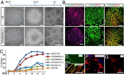
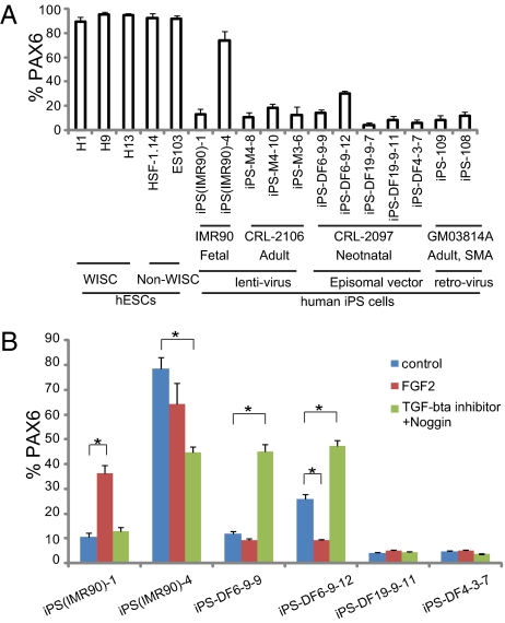
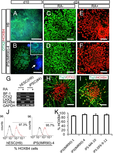
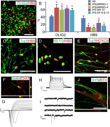
For the promise of human induced pluripotent stem cells (iPSCs) to be realized, it is necessary to ask if and how efficiently they may be differentiated to functional cells of various lineages. Here, we have directly compared the neural-differentiation capacity of human iPSCs and embryonic stem cells (ESCs). We have shown that human iPSCs use the same transcriptional network to generate neuroepithelia and functionally appropriate neuronal types over the same developmental time course as hESCs in response to the same set of morphogens; however, they do it with significantly reduced efficiency and increased variability. These results were consistent across iPSC lines and independent of the set of reprogramming transgenes used to derive iPSCs as well as the presence or absence of reprogramming transgenes in iPSCs. These findings, which show a need for improving differentiation potency of iPSCs, suggest the possibility of employing human iPSCs in pathological studies, therapeutic screening, and autologous cell transplantation.


Conflict of interest statement

Conflict of interest statement: J.A.T. is a founder, stock owner, consultant, and board member of Cellular Dynamics International (CDI). He also serves as a scientific advisor to and has financial interests in Tactics II Stem Cell Ventures.

Figures

Fig. 1.
Fig. 1.
Human iPSCs and hESCs follow the same temporal course of neural differentiation. (A) Phase contrast images show that hESCs and iPSCs grew as individual colonies, differentiated to columnar epithelial cells at days 8–10, and formed neural tube-like rosettes at day 15. (B) Both iPSCs and hESCs were positive for OCT4 at day 0, for PAX6 but not SOX1 at days 8–10, and for both PAX6 and SOX1 at day 15. (C) FACS analyses indicate that differentiating cells from H9 hESCs, iPS(IMR90)-1 and 4, iPS-M4-10, iPS-DF6-9–12, and iPS109 began to generate PAX6-expressing cells at days 6–8, and this reached a plateau at day 14 but with different efficiency. Shown are curves of the average from three replicates for each cell line. (D and E) By 12 weeks in culture, many MAP2+ neurons also expressed synapsin; higher magnification indicated a punctuate staining pattern on the cell bodies and neurites. (F) GFAP+ astrocytes were present in differentiated cultures at 12 weeks. (G) O4+ ramified oligodendrocytes were observed in cultures after 16 weeks. Except when noted elsewhere, images of iPSCs are presented with iPS(IMR90)-4 in this and the following figures. (Scale bar, 50 μm.)
Fig. 2.
Fig. 2.
Variation of iPSC neural differentiation. (A) At day 15, FACS analyses revealed a similar proportion of PAX6-expressing cells from five hESC lines but variable percentages for the iPSC lines. (B) The human iPSC lines responded to FGF2 or dual SMAD inhibition variably. Data are shown as mean ± SEM (n = 3). Asterisk denotes P < 0.001.
Fig. 3.
Fig. 3.
iPSC-derived neuroepithelial cells can be patterned to regional progenitors and differentiated to neurons and glia. (A and B) Human iPSC- and ESC-derived neuroepithelial cells were positive for OTX2 and negative for HOXB4 at day 10. Some cell clusters (Hoechst labeled; arrows) in the iPSC cultures were negative for OTX2. Most of the OTX2+ cluster formed typical rosettes 4 days later as shown in B Inset. (C–F) By day 24, cultures treated with RA were now positive for HOXB4 but negative for OTX2, whereas those without RA treatment remained OTX2 positive. (G) RT-PCR showed expression of BF-1, OTX2, and LHX2 but not HOXB4 in neuroepithelial cultures without RA treatment and with HOXB4 expression in RA-treated neuroepithelia at day 24. (H) Further differentiation shows that without RA, iPSC-derived βIII-tubulin+ neurons were positive for OTX2 but were labeled for HOXB4 in the presence of RA (I). (J) FACS showed that the majority of hESC- and iPSC-derived neuroepithelia was positive for HOXB4 in response to RA treatment. (K) For the representative iPS cell lines tested, more than 90% of the neural epithelial cells could be caudalized to HOXB4-expressing progenitors.
Fig. 4.
Fig. 4.
iPSCs differentiate to functional motor neurons. (A) iPSC-derived neural cells expressed OLIG2 and HB9 at day 35. (B) FACS analysis of HB9+ motor neurons differentiated from hESCs and iPSCs. Data are shown as mean ± SEM (n = 3). Asterisk denotes P < 0.01 by Dunnett's test with H9 as a reference. (C) HB9 was expressed on differentiation to TuJ1+ neuronal cells. (D) iPSC-derived HB9+ cells were positive for ISL1/2 shown by a single confocal section. (E) Confocal imaging shows HOXC8 staining in the nuclei of iPSC-derived Tuj1+ neurons. (F) Cells targeted for physiological analysis were labeled with neurobiotin and stained for ChAT+ to verify their motor neuron (MN) identity. (G) I-Clamp traces revealed multiple APs generated during 500-ms current injections of various amplitudes. (H) Expanded view of step-induced currents revealed large, rapidly inactivating inward currents followed by sustained outward currents. (I) Example traces of spontaneous postsynaptic currents detected in iPSC-derived MNs. (J) Confocal imaging showed that BTX was localized to the area where synapsin+ neurites contacted the myotube. (Scale bar, 50 μm.)

References

    1. Perrier AL, et al. Derivation of midbrain dopamine neurons from human embryonic stem cells. Proc Natl Acad Sci USA. 2004;101:12543–12548. - PMC - PubMed
    1. Li XJ, et al. Specification of motoneurons from human embryonic stem cells. Nat Biotechnol. 2005;23:215–221. - PubMed
    1. Roy NS, et al. Functional engraftment of human ES cell-derived dopaminergic neurons enriched by coculture with telomerase-immortalized midbrain astrocytes. Nat Med. 2006;12:1259–1268. - PubMed
    1. Watanabe K, et al. A ROCK inhibitor permits survival of dissociated human embryonic stem cells. Nat Biotechnol. 2007;25:681–686. - PubMed
    1. Yang L, et al. Human cardiovascular progenitor cells develop from a KDR+ embryonic-stem-cell-derived population. Nature. 2008;453:524–528. - PubMed

Publication types

Substances

LinkOut - more resources