. 2010 Mar;24(3):574-86.
doi: 10.1210/me.2009-0369. Epub 2010 Feb 16.

Agonist-induced endocytosis and receptor phosphorylation mediate resensitization of dopamine D(2) receptors

Affiliations

Agonist-induced endocytosis and receptor phosphorylation mediate resensitization of dopamine D(2) receptors

Dongim Cho et al. Mol Endocrinol. 2010 Mar.

Abstract

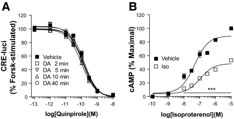
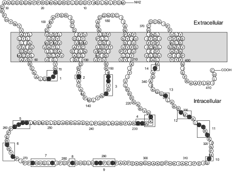
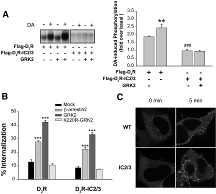
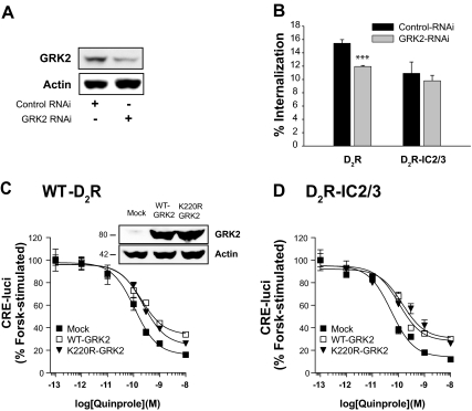
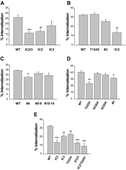
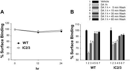
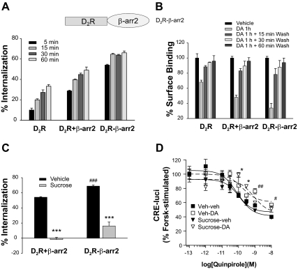
The regulatory mechanisms and functional roles of agonist-induced internalization of G protein-coupled receptors (GPCRs) were analyzed using mutant dopamine D(2) receptors (D(2)Rs) in which all possible GPCR kinase (GRK) phosphorylation sites were mutated or the affinity for beta-arrestins was altered. Agonist-induced internalization of D(2)Rs involved a phosphorylation-dependent component, which was mediated by serine/threonine (S/T) residues in the second loop and T225 in the third loop, and a phosphorylation-independent component. GRK2-mediated enhancement of the internalization and inhibition of D(2)R signaling did not involve receptor phosphorylation, and only the former required the enzymatic activity of GRK2. The phosphorylation-deficient mutant (D(2)R-intracellular loop 2/3) recycled more slowly and showed more agonist-induced desensitization than did the wild-type D(2)R, suggesting that receptor phosphorylation mediates the recycling of the internalized receptors and enhances receptor resensitization. Blockade of the agonist-induced internalization of D(2)R-intracellular loop 2/3 provoked desensitization as in wild-type D(2)R, suggesting that certain cellular processes other than receptor dephosphorylation occurring within the endocytic vesicle are responsible for the resensitization of D(2)R. When dissociation between D(2)R and beta-arrestin was inhibited or when the expression of cellular beta-arrestins was decreased, agonist-induced desensitization of D(2)R did not occur, suggesting that dissociation from beta-arrestin is the main cellular process required for resensitization of D(2)R and is achieved through agonist-induced internalization. These results indicate that, in the regulation of some GPCRs, phosphorylation-independent association with beta-arrestin plays a major role in agonist-induced desensitization.


Figures

Figure 1
Figure 1
Characterization of homologous desensitization of the D2R. A, Agonist-induced desensitization of the D2R. HEK-293 cells expressing D2R were pretreated with either serum-free medium containing 100 μm ascorbic acid (vehicle) or 10 μm DA dissolved in the vehicle for 2–40 min. Cellular cAMP was measured using CRE-luci reporter gene as described in Materials and Methods. Each data point represents the mean ± sem. Forsk, Forskolin. B, Agonist-induced desensitization of β2AR. Cells expressing β2AR were pretreated with 10 μm isoproterenol, and the cAMP was measured as described in Materials and Methods. ***, P < 0.001 compared with the vehicle group at the same concentration of isoproterenol. The levels of cAMP produced by the stimulation of β2AR after preexposure to isoproterenol were significantly lower than levels in the vehicle group (P < 0.001) at concentrations of isoproterenol higher than 10−8 m. Iso, Isoproterenol.
Figure 2
Figure 2
Diagram of the D2R mutants and their putative phosphorylation sites. The shaded region represents the putative transmembrane region. Site-directed mutagenesis was performed to change serine (S) or threonine (T) residues designated within the cytoplasmic loops (nos. 1–14) into alanine or valine residues, respectively. Detailed information on the mutants is given in Table 1.
Figure 3
Figure 3
Characterization of phosphorylation-dependent and phosphorylation-independent internalization of the D2R. A, Agonist-induced phosphorylation of D2R mutants. HEK-293 cells were transiently transfected with Flag-D2R or Flag-D2R-IC2/3 with or without GRK2-pRK5. Receptor phosphorylation was determined after 5 min of stimulation with 10 μm DA. Receptor expression was measured by radioligand binding, and the equal amount of receptor protein was loaded in each lane. The phosphorylated receptors were visualized by autoradiography. **, P < 0.01 when the Flag-D2R/GRK2 group was compared with the Flag-D2R/Mock group. ###, P < 0.001 when the Flag-D2R-IC2/3 group was compared with the Flag-D2R group. B, Effects of GRK2 and β-arrestins on phosphorylation-dependent and phosphorylation-independent internalization of the D2R. WT-D2R or D2R-IC2/3 was cotransfected with βarr2-pCMV5 (2 μg), GRK2-pRK5 (2 μg), or K220R-GRK2-pRK5 (2 μg). Receptor expression levels were maintained around 1.2 pmol/mg protein. Receptor internalization was determined as described in Materials and Methods. ***, P < 0.001 compared with the Mock group. C, β-Arrestin translocation assay. HEK-293 cells were transfected with 3 μg of the corresponding receptor constructs and 1.5 μg βarr2-GFP per 100-mm culture dish. Cells were stimulated with 10 μm DA for 5 min. Receptor expression levels were measured using [3H]spiperone at 3 nm and maintained at approximately 3 pmol/mg protein. β-Arrestin translocation was not observed in HEK-293 cells that were not tranfected with D2R (Supplemental Fig. 1A). Similar extent of β-arrestin translocation was observed in WT-D2R and D2R-IC2/3 at 1-min treatment with DA (Supplemental Fig. 1, B and C). βarr2, β-arrestin 2.
Figure 4
Figure 4
Roles of GRK2 in the internalization and signaling of D2R. A, Inhibition of GRK2 expression by RNAi. Cells were transfected with RNAi plasmids of GFP [in pcDNA3.1/Zeo(+)] and of GRK2 [pcDNA3.1/Zeo(+)], and stably transfected cells were selected. Cellular GRK2 was detected by immunoblotting using antibodies against GRK2. B, Effects of knockdown of GRK2 on the internalization of D2R. Cells were transfected with D2R-pCMV5 (∼1.3 pmol/mg protein), and internalization was determined using [3H]sulpiride binding. ***, P < 0.001 compared with Control-RNAi group. C and D, Effects of GRK2 on the signaling of D2R. Cells were transfected with 2 μg WT- or K220R-GRK2 together with WT-D2R (C) or D2R-IC2/3 (D). Cellular cAMP was measured using CRE-luci reporter gene as described in Materials and Methods. P < 0.01–0.001 when the Mock group was compared with the WT-GRK2 group or K220R-GRK2 group at doses of quinpirole between 3 × 10⁻¹⁰ and 10−8 m (C), and between 10−10 and 10−8 m (D) except that not statistically significant between Mock and WT-GRK2 group at 3 × 10⁻¹⁰ m. Expression levels of WT-GRK2 and K220R-GRK2 were determined by immunoblotting the cell lysates with antibodies to GRK2.
Figure 5
Figure 5
Determination of the serine and threonine residues responsible for agonist-induced internalization of the D2R. HEK-293 cells transfected with each mutant D2R and βarr2 were treated with 10 μm DA for 1 h. Receptor expression levels were maintained around 1.8 pmol/mg protein. *, P < 0.05; **, P < 0.01; ***, P < 0.001 compared with WT-D2R.
Figure 6
Figure 6
Functional roles of agonist-induced internalization of D2R. A, Inhibition of D2R internalization by sucrose. Cells were treated with 0.45 m sucrose for 20 min, washed with serum-free medium three times for 5 min per wash, and then treated with 10 μm DA for 1 h. ***, P < 0.001 compared with each vehicle-treated group. B, Effects of the inhibition of agonist-induced internalization on the homologous desensitization of WT-D2R. The desensitization assay was conducted before and after sucrose treatment. **, P < 0.01; ***, P < 0.001 when the sucrose/DA group was compared with other experimental groups (Veh-veh, Sucrose-veh, Veh-DA). C, Agonist-induced desensitization of the phosphorylation-independent mutant of D2R. Desensitization studies were conducted for D2R-IC2/3 before and after sucrose treatment. P < 0.01 when the vehicle-DA group was compared with the vehicle-vehicle group or sucrose-vehicle group at doses of quinpirole between 10−10 and 10−8 m. P < 0.01 when sucrose-DA group was compared with vehicle-DA group. P < 0.001 when sucrose-DA group was compared with vehicle-vehicle or sucrose-vehicle group. veh, Vehicle.
Figure 7
Figure 7
Characterization of the intracellular trafficking of phosphorylation-independent mutants of the D2R. A, Comparisons of agonist-induced down-regulation of D2R. HEK-293 cells that stably expressed WT-D2R or D2R-IC2/3 were treated with 1 μm quinpirole for 0, 12, and 24 h. Cells were washed three times with ice-cold serum-free medium for 10 min each, and ligand-binding studies were conducted at 4 C for 150 min. B, Comparisons of agonist-induced internalization and recycling of D2R. HEK-293 cells that stably expressed WT-D2R or D2R-IC2/3 were treated with 10 μm DA for 1 h. After washing with serum-free medium, cells were incubated at 37 C for the indicated period of time.
Figure 8
Figure 8
Roles of βarr2 on sucrose/agonist-induced desensitization of D2R. A, Inhibition of β-arrestin expression by RNAi plasmids of β-arrestin1 [pcDNA3.1/Zeo(+)] and βarr2 [pcDNA3.0 (Neo)]. Stably transfected cells were selected, and the cellular β-arrestin was detected by immunoblotting using antibodies against β-arrestin1 or βarr2. B, Effects of sucrose treatment on the agonist-induced internalization of D2R in control and βarr2 RNAi cells. Cells were treated as in Fig. 6A. C and D, Effects of knockdown of cellular βarr2 on agonist-induced desensitization of the WT-D2R (C) and D2R-IC2/3 (D). E and F, Effects of exogenous β-arrestin2 on agonist-induced desensitization of the WT-D2R (E) and D2R-IC2/3 (F) in βarr2 knockdown cells. *, P < 0.05; **, P < 0.01, ***, P < 0.001 when sucrose-DA group was compared with other experimental groups. #, P < 0.05; ##, P < 0.01 when vehicle-DA group was compared with vehicle-vehicle or sucrose-vehicle group. veh, Vehicle.
Figure 9
Figure 9
Effects of increased affinity between D2R and βarr2 on agonist-induced desensitization of the D2R. A, Effects of fusion of βarr2 to the D2R on agonist-induced internalization. Receptor expression levels were maintained around 1 pmol/mg protein. B, Effects of fusion of βarr2 to D2R on recycling of internalized receptors. P < 0.001 when each DA 1 h group was compared with corresponding vehicle group. P < 0.01 (D2R +βarr2) and P < 0.05 (D2R-βarr2) when each DA 1 h + 15 min wash group was compared with corresponding vehicle group. C, Effects of sucrose treatment on agonist-induced internalization of WT-D2R and D2R-βarr2. Cells were treated as in Fig. 6A. ***, P < 0.001 when compared with each vehicle group. ###, P < 0.001 when compared with D2R +βarr2, vehicle group. D, Effects of fusion of βarr2 to D2R on agonist-induced desensitization. Cells were treated as in Fig. 6B. *, P < 0.05 when sucrose-DA group is compared with vehicle-vehicle or sucrose-vehicle group. #, P < 0.05; ##, P < 0.01 when vehicle-DA group is compared with vehicle-vehicle or sucrose-vehicle group.

Similar articles

Cited by

References

    1. Lohse MJ, Lefkowitz RJ, Caron MG, Benovic JL 1989 Inhibition of β-adrenergic receptor kinase prevents rapid homologous desensitization of β 2-adrenergic receptors. Proc Natl Acad Sci USA 86:3011–3015 - PMC - PubMed
    1. Hausdorff WP, Caron MG, Lefkowitz RJ 1990 Turning off the signal: desensitization of β-adrenergic receptor function. FASEB J 4:2881–2889 - PubMed
    1. Gurevich VV, Benovic JL 1997 Mechanism of phosphorylation-recognition by visual arrestin and the transition of arrestin into a high affinity binding state. Mol Pharmacol 51:161–169 - PubMed
    1. Sibley DR, Strasser RH, Benovic JL, Daniel K, Lefkowitz RJ 1986 Phosphorylation/dephosphorylation of the β-adrenergic receptor regulates its functional coupling to adenylate cyclase and subcellular distribution. Proc Natl Acad Sci USA 83:9408–9412 - PMC - PubMed
    1. Ferguson SS, Downey III WE, Colapietro AM, Barak LS, Ménard L, Caron MG 1996 Role of β-arrestin in mediating agonist-promoted G protein-coupled receptor internalization. Science 271:363–366 - PubMed

Publication types

MeSH terms