. 2010 Mar;24(3):608-20.
doi: 10.1210/me.2008-0374. Epub 2010 Feb 16.

Inhibin-A antagonizes TGFbeta2 signaling by down-regulating cell surface expression of the TGFbeta coreceptor betaglycan

Affiliations

Inhibin-A antagonizes TGFbeta2 signaling by down-regulating cell surface expression of the TGFbeta coreceptor betaglycan

Brendan D Looyenga et al. Mol Endocrinol. 2010 Mar.

Abstract

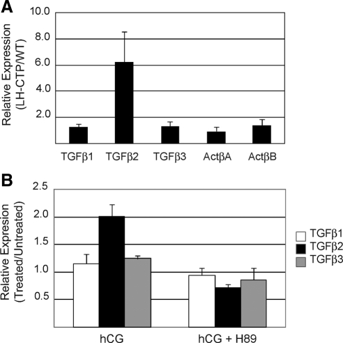
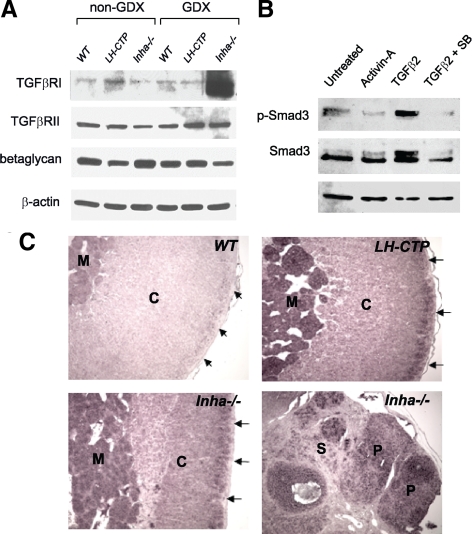
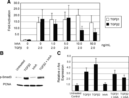
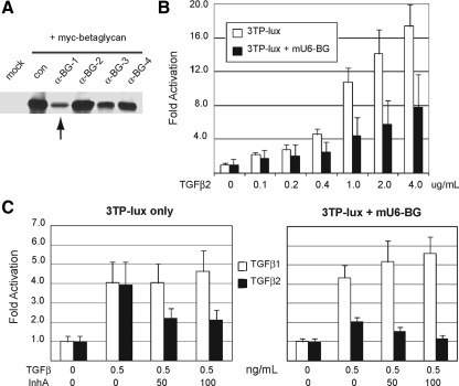
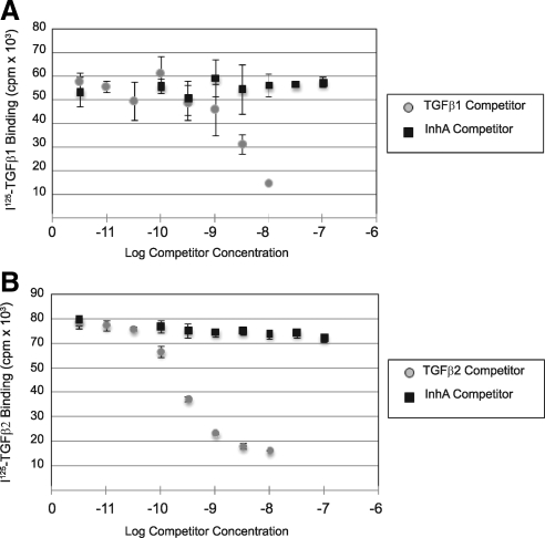
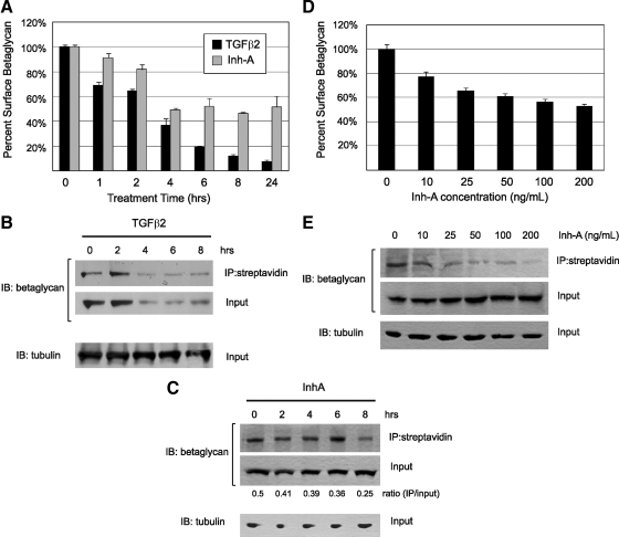
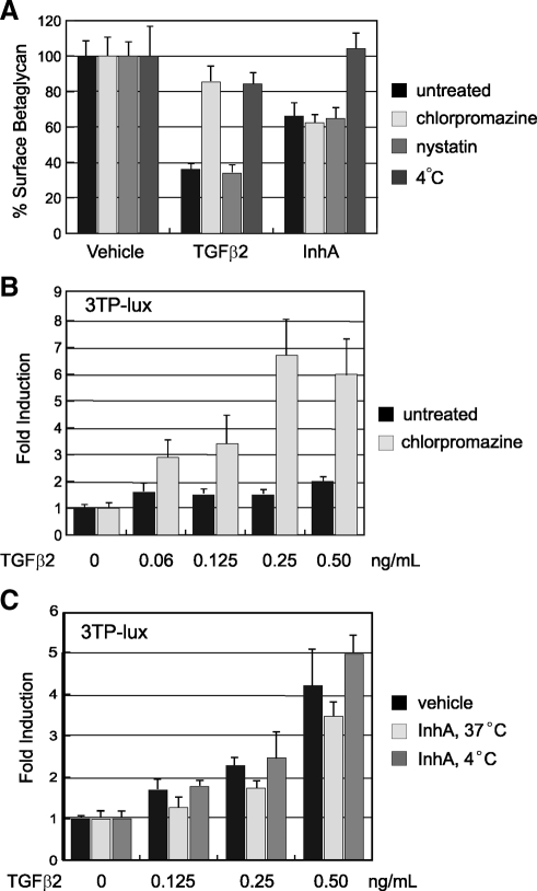
Inhibin is an atypical member of the TGFbeta family of signaling ligands and is classically understood to function via competitive antagonism of activin ligand binding. Inhibin-null (Inha-/-) mice develop both gonadal and adrenocortical tumors, the latter of which depend upon gonadectomy for initiation. We have previously shown that gonadectomy initiates adrenal tumorigenesis in Inha-/- mice by elevating production of LH, which drives aberrant proliferation and differentiation of subcapsular adrenocortical progenitor cells. In this study, we demonstrate that LH signaling specifically up-regulates expression of TGFbeta2 in the subcapsular region of the adrenal cortex, which coincides with regions of aberrant Smad3 activation in Inha-/- adrenal glands. Consistent with a functional interaction between inhibin and TGFbeta2, we further demonstrate that recombinant inhibin-A antagonizes signaling by TGFbeta2 in cultured adrenocortical cells. The mechanism of this antagonism depends upon the mutual affinity of inhibin-A and TGFbeta2 for the signaling coreceptor betaglycan. Although inhibin-A cannot physically displace TGFbeta2 from its binding sites on betaglycan, binding of inhibin-A to the cell surface causes endocytic internalization of betaglycan, thereby reducing the number of available binding sites for TGFbeta2 on the cell surface. The mechanism by which inhibin-A induces betaglycan internalization is clathrin independent, making it distinct from the mechanism by which TGFbeta ligands themselves induce betaglycan internalization. These data indicate that inhibin can specifically antagonize TGFbeta2 signaling in cellular contexts where surface expression of betaglycan is limiting and provide a novel mechanism for activin-independent phenotypes in Inha-/- mice.


Figures

Figure 1
Figure 1
TGFβ2 is specifically up-regulated by LHR signaling. A, Quantitative RT-PCR analysis of TGFβ and activin subunit gene expression was performed on RNA transcripts harvested from the adrenal glands of wild-type and bLH-βCTP mice. Values for each transcript were normalized to Arbp expression and are displayed as the ratio of average values from bLH-βCTP mice to wild-type mice. Error bars represent the standard deviation of seven mice per group. B, Primary Inha−/− adrenocortical tumor cells were explanted to culture and treated 24 h with media only, hCG (50 ng/ml), or hCG and the PKA inhibitor H89 (2 μm). Quantitative RT-PCR analysis was performed on RNA harvested from each sample. Values for each gene were normalized to 18s rRNA expression and are displayed as the ratio of values from treated and untreated cells. Error bars represent the standard deviation of three separate experimental replicates. WT, Wild type.
Figure 2
Figure 2
TGFβ2 is expressed in the adrenal cortex and activates Smad3 signaling in primary mouse adrenocortical cells. A, Immunoblot analysis of TGFβ receptor expression was performed on 10–40 μg of whole adrenal gland protein lysates from 2 month-old female mice of the indicated genotype. Equal protein amounts were loaded for each sample. Blots were probed with antibodies to TGFβRI (Alk5), TGFβRII, and TGFβRIII (betaglycan), as well as β-actin to demonstrate equal protein loading. B, Adrenals from six age-matched wild-type female mice were pooled and explanted to primary culture in six-well plates. After 16 h of culture, the cells were washed and treated with vehicle alone (PBS, lane1), activin-A (2 ng/ml, lane 2), TGFβ2 (1 ng/ml, lane 3), or TGFβ2 plus the Alk5 inhibitor SB431542 (1 μm, lane 4) for 16 h. Protein lysates were loaded in equal amounts for each sample and submitted to immunoblot analysis for expression and activation (phosphorylation) of Smad3. Blots were also probed for β-actin to indicate equal protein loading. C, Nonradioactive in situ hybridization analysis of TGFβ2 expression was performed on sections of paraffin-embedded adrenal glands harvested from wild-type (WT), bLH-βCTP, and Inha−/− mice 20–30 wk after gonadectomy. Regions of the tissue that express TGFβ2 transcripts hybridize to the digoxigenin-labeled RNA probe and are detected by alkaline phosphatase activity (purple) produced by an anti-DIG/AP-conjugated antibody. The bottom two panels are derived from adjacent normal adrenal and adrenocortical tumor tissue from the same Inha−/− adrenal sample. Staining in the adrenal cortex (C) is enhanced in the subcapsular region (arrows) and is also prominently seen in tumor parenchyma (P), but not in the cortical stroma (S). The adrenal medulla (M) also stains purple due to endogenous alkaline phosphatase activity. AP, Alkaline phosphatase; GDX, gonadectomy; DIG, digoxigenin.
Figure 3
Figure 3
Inhibin functionally antagonizes TGFβ2 signaling in vitro. A, Luminescent assays for 3TP-lux reporter activation were performed in Y1 adrenocortical cells treated with a fixed amount of TGFβ1 or TGFβ2 (2 ng/ml) and increasing doses of recombinant inhibin-A peptide as indicated. 3TP-lux activity was normalized to the luminescent signal from a constitutively expressed pCMV-Renilla reporter vector that was cotransfected with 3TP-lux into Y1 cells. B, Y1 cells were treated with vehicle (PBS), TGFβ2 (0.5 ng/ml), inhibin-A (100 ng/ml), or TGFβ2 plus inhibin-A for 16 h. Immunoblot analysis was performed on 10–20 μg of protein lysates loaded in equal amounts for each sample. Blots were probed with antibodies to phospho-Smad3 to indicate TGFβ2 signaling activity and proliferating cell nuclear antigen to indicate equal protein loading. C, Y1 cells were treated with vehicle (PBS), TGFβ1 (0.5 ng/ml), TGFβ2 (0.5 ng/ml), inhibin-A (100 ng/ml), or TGFβ ligand plus inhibin-A for 16 h. Quantitative RT-PCR analysis of c-fos gene expression was performed on total RNA extracted from each sample. Values for c-fos were normalized to Arbp expression, with error bars indicating the standard error of three separate measurements. PCNA, Proliferating cell nuclear antigen.
Figure 4
Figure 4
TGFβ2 signaling and inhibin antagonism depend on betaglycan expression. A, 293-HEK cells were transfected with pCDNA3-betaglycan-myc alone or with one of four mU6-BG shRNA targeting vectors to validate the effectiveness of hairpin RNAs in decreasing betaglycan expression. Immunoblot analysis was used to indicate myc-betalycan expression in cells after cotransfection. Vector mU6-BG1 (lane 3, arrow) was the most effective at decreasing betaglycan expression and was used in subsequent assays. B, Luminescent assays for 3TP-lux reporter activation were performed in Y1 adrenocortical cells treated with increasing concentrations of TGFβ2 with or without the mU6-BG vector. C, Luminescent assays for 3TP-lux reporter activation were performed in Y1 adrenocortical cells treated with TGFβ1 or TGFβ2 either alone or in combination with recombinant inhibin-A. Experiments were repeated with cotransfection of the mU6-BG targeting vector to evaluate the effect of decreased betaglycan expression on signaling efficiency. For all reporter assays, 3TP-lux activity was normalized to signal from a CMV-Renilla luciferase cotransfected with 3TP-lux into Y1 cells. Data shown are triplicate measurements with error bars indicating standard deviations from the average of these three values. BG, Beta glycan.
Figure 5
Figure 5
Inhibin-A cannot physically displace TGFβ ligands from cell surface-binding sites. Binding assays with [125I]TGF-β1 and [125I]TGF-β2 were performed on intact Y1 cells in 12-well plates. A, Binding of [125I]TGF-β1 to Y1 cells was competed with the indicated concentrations of unlabeled TGFβ1 ligand and unlabeled recombinant inhibin-A. B, Binding of [125I]TGF-β2 was similarly competed with the indicated concentrations of unlabeled TGFβ1 ligand and unlabeled recombinant inhibin-A.
Figure 6
Figure 6
Inhibin-A induces betaglycan internalization. A, Y1 cells were treated with recombinant TGFβ2 (2 ng/ml) or inhibin-A (NIBSC, 100 ng/ml) in serum-free DMEM containing 0.05% BSA for the indicated times. Intact/live cells were labeled with an antibetaglycan antibody and analyzed by flow cytometry. Mean fluorescent signal strength was determined for each cell population. Signal from intact cells in each treated sample was normalized against signal from untreated Y1 cells, which was considered 100% surface occupation. Experiments were performed in triplicate, and data are displayed with error bars representing the standard deviations from these three replicate measurements. B, Surface proteins from Y1 cells treated for the indicated times with 2 ng/ml TGFβ2 were biotinylated and precipitated with streptavidin beads. Immunoblots for betaglycan and tubulin were performed on equal amounts of precipitate and 10% of the input lysates for each sample. C, Y1 cells treated for the indicated times with 100 ng/ml inhibin-A were biotinylated and treated as indicated above. Immunoblots for betaglycan and tubulin were performed on equal amounts of precipitate and 10% of the input lysates for each sample. Protein quantity was determined by densitometric analysis of band intensity, and the ratio of surface to total betaglycan is displayed beneath the lane of each experimental condition. D, Live/intact Y1 cells treated for 6 h with the indicated concentration of inhibin-A were labeled for surface betaglycan and analyzed by flow cytometry. Values shown were normalized as indicated above, with error bars representing standard deviation for three independent replicates. E, Y1 cells treated for 6 h with the indicated concentration of inhibin-A were biotinylated and treated as indicated above. Immunoblots for betaglycan and tubulin were performed on equal amounts of precipitate and 10% of the input lysates for each sample. IB, Immunoblot; IP, immunoprecipitation.
Figure 7
Figure 7
Inhibin-A promotes endocytosis of betaglycan by a clathrin-independent mechanism. A, Y1 cells were treated with recombinant TGFβ2 (2 ng/ml) or inhibin-A (NIBSC, 100 ng/ml) in serum-free DMEM containing chlorpromazine (25 μm), nystatin (25 μg/ml), or incubation at 4 C for 6 h. Intact/live cells were labeled with an antibetaglycan antibody and analyzed by flow cytometry. Mean fluorescent signal strength was determined for each cell population, and was normalized against the value from untreated Y1 cells, which was considered 100% surface occupation. Experiments were performed in triplicate and data is displayed with error bars representing the standard deviations from these three replicate measurements. B, Luminescent assays for 3TP-lux reporter activation were performed in Y1 adrenocortical cells treated for 16 h with 25 μm chlorpromazine and the indicated concentration of TGFβ2. 3TP-lux activity was normalized to the luminescent signal from a constitutively expressed pCMV-Renilla reporter vector that was cotransfected with 3TP-lux into Y1 cells. Values for three independent replicates are shown with error bars indicated standard deviation. C, Luminescent assays for 3TP-lux reporter activation were performed in Y1 adrenocortical cells treated for 8 h with 100 ng/ml recombinant inhibin-A at either 37 C or 4 C, followed by 16 h with the indicated concentration of TGFβ2 at 37 C. 3TP-lux activity was normalized as above, with error bars indicated standard deviation.

Similar articles

Cited by

References

    1. Chang H, Brown CW, Matzuk MM 2002 Genetic analysis of the mammalian transforming growth factor-β superfamily. Endocr Rev 23:787–823 - PubMed
    1. Matzuk MM, Lamb DJ 2002 Genetic dissection of mammalian fertility pathways. Nat Cell Biol 4(Suppl):s41–s49 - PubMed
    1. Cook RW, Thompson TB, Jardetzky TS, Woodruff TK 2004 Molecular biology of inhibin action. Semin Reprod Med 22:269–276 - PubMed
    1. Ethier JF, Findlay JK 2001 Roles of activin and its signal transduction mechanisms in reproductive tissues. Reproduction 121:667–675 - PubMed
    1. Massagué J, Seoane J, Wotton D 2005 Smad transcription factors. Genes Dev 19:2783–2810 - PubMed

Publication types

MeSH terms