. 2010 Mar;120(3):713-9.
doi: 10.1172/JCI41366. Epub 2010 Feb 15.

Elevated Tribbles homolog 2-specific antibody levels in narcolepsy patients

Affiliations

Elevated Tribbles homolog 2-specific antibody levels in narcolepsy patients

Vesna Cvetkovic-Lopes et al. J Clin Invest. 2010 Mar.

Abstract

Narcolepsy is a sleep disorder characterized by excessive daytime sleepiness and attacks of muscle atonia triggered by strong emotions (cataplexy). Narcolepsy is caused by hypocretin (orexin) deficiency, paralleled by a dramatic loss in hypothalamic hypocretin-producing neurons. It is believed that narcolepsy is an autoimmune disorder, although definitive proof of this, such as the presence of autoantibodies, is still lacking. We engineered a transgenic mouse model to identify peptides enriched within hypocretin-producing neurons that could serve as potential autoimmune targets. Initial analysis indicated that the transcript encoding Tribbles homolog 2 (Trib2), previously identified as an autoantigen in autoimmune uveitis, was enriched in hypocretin neurons in these mice. ELISA analysis showed that sera from narcolepsy patients with cataplexy had higher Trib2-specific antibody titers compared with either normal controls or patients with idiopathic hypersomnia, multiple sclerosis, or other inflammatory neurological disorders. Trib2-specific antibody titers were highest early after narcolepsy onset, sharply decreased within 2-3 years, and then stabilized at levels substantially higher than that of controls for up to 30 years. High Trib2-specific antibody titers correlated with the severity of cataplexy. Serum of a patient showed specific immunoreactivity with over 86% of hypocretin neurons in the mouse hypothalamus. Thus, we have identified reactive autoantibodies in human narcolepsy, providing evidence that narcolepsy is an autoimmune disorder.


Figures
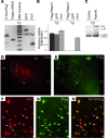
Figure 1
Figure 1. Pabpc1-hypocretin transgenic mice.
(A) RT-PCR experiments performed on total RNAs from a transgenic mouse hypothalamus (Tg+), showing the presence of both the construct and hypocretin (Hcrt) transcripts. Four different primer pairs were used, each generating a PCR fragment of the size predicted by its cDNA sequence: 5′ Hcrt/Flag (467 bp), Pabpc1/IRES (333 bp), GFP (611 bp), and Hcrt (189 bp). (B) Quantitative RT-PCR experiments showing similar relative RNA expression levels for Flag-tagged Pabpc1, GFP, and Hcrt in transgenic mice hypothalamus. Only the Hcrt transcript was detected in nontransgenic (Tg-) mouse hypothalamus as expected. The normalized relative values (+ 1 SD) obtained correspond to the mean values from 4 biological replicates per condition. (C) Western blot analysis demonstrating the presence of the Flag protein in the hypothalamic (hypoth.) extract (obtained from 3 transgenic mice) and its absence in the cortical fraction (from the same 3 mice). The loading control is a nonspecific band generated by the mouse monoclonal anti-Flag M2 antibody. Photomicrographs illustrating the distribution of (D and F) Hcrt- and (E and G) Flag-expressing cells on a coronal section from the tuberal hypothalamus. (FH) Higher magnification views of the dashed regions in D and E confirm that Hcrt and Flag are coexpressed in the same neurons. Arrows indicate both single Hcrt or Flag or double Hcrt-Flag staining. Almost all Hcrt neurons were found to express the Flag-containing construct. (H) Merged image of F and G. cp, cerebral peduncle; fx, fornix; mtt, mammilo-thalamic tract; 3V, third ventricle. Scale bar: 100 μm (D and E); 20 μm (FH).
Figure 2
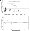
Figure 2. ELISA determination of Trib2-specific antibodies in sera.
(A) Each symbol corresponds to the serum of a single subject. Mean ± 1 SD of each group is shown next to the individual values. The dotted horizontal line indicates the mean Trib2-specific antibody titer in healthy control subjects plus 2 SD. All values are relative to the optical density of a healthy control subject (which is equal to 1). P values correspond to independent t-tests between indicated groups. OIND, other inflammatory neurological diseases. (B) Mean ± 1 SD of Trib2-specific antibody titers at different intervals from the disease onset. The solid line and dotted lines indicate mean titer ± 1 SD in normal control subjects. Numbers indicate the number of narcolepsy patients at each interval. Note the sharp decrease in titers within the first 2–3 years, reaching normal values. From 5 up to 30 years after disease onset, the titers of narcolepsy patients remain stable but significantly (1 SD, P < 5 × 10⁻⁵) higher than those of healthy control subjects (n = 42).
Figure 3
Figure 3. Double immunohistochemistry with hypocretin antibody and a high Trib2-specific antibody-containing serum of a narcolepsy with cataplexy patient.
(A) Coronal section through the mouse hypothalamus, stained with anti-hypocretin antibody. (B) The same section stained with the patient’s serum. (C) Merged hypocretin-serum staining showing that the vast majority of hypocretin-positive neurons are costained by the patient’s serum. (DF) Higher magnification views of the dashed regions in AC confirm the double labeling of hypocretin neurons. Overall, 87% of hypocretin neurons are costained by the serum. (G) Another mouse hypothalamic section stained with anti-hypocretin antibody. (H) Immunoreactivity with the same patient’s serum as in B but after preabsorption of Trib2-specific antibodies with excess Trib2 peptide. (I) Merged image showing that the vast majority of hypocretin neurons are only stained with hypocretin antibody. Only 8% of hypocretin neurons are double labeled. (JL) Higher magnification views of the dashed regions in GI confirm that serum depletion of Trib2-specific antibodies results in the absence of double staining. Scale bar: 20 μm.

Comment in

Similar articles

Cited by

References

    1. Dauvilliers Y, Arnulf I, Mignot E. Narcolepsy with cataplexy. Lancet. 2007;369(9560):499–511. doi: 10.1016/S0140-6736(07)60237-2. - DOI - PubMed
    1. Thannickal TC, et al. Reduced number of hypocretin neurons in human narcolepsy. Neuron. 2000;27(3):469–474. doi: 10.1016/S0896-6273(00)00058-1. - DOI - PMC - PubMed
    1. Peyron C, et al. A mutation in a case of early onset narcolepsy and a generalized absence of hypocretin peptides in human narcoleptic brains. Nat Med. 2000;6(9):991–997. doi: 10.1038/79690. - DOI - PubMed
    1. Willie JT, et al. Distinct narcolepsy syndromes in Orexin receptor-2 and Orexin null mice: molecular genetic dissection of Non-REM and REM sleep regulatory processes. Neuron. 2003;38(5):715–730. doi: 10.1016/S0896-6273(03)00330-1. - DOI - PubMed
    1. Hara J, et al. Genetic ablation of orexin neurons in mice results in narcolepsy, hypophagia, and obesity. Neuron. 2001;30(2):345–354. doi: 10.1016/S0896-6273(01)00293-8. - DOI - PubMed

Publication types

MeSH terms