

Role of peroxisome proliferator-activated receptor-alpha in hepatobiliary injury induced by ammonium perfluorooctanoate in mouse liver
- PMID: 20160413
- PMCID: PMC7385711
- DOI: 10.2486/indhealth.48.96
Role of peroxisome proliferator-activated receptor-alpha in hepatobiliary injury induced by ammonium perfluorooctanoate in mouse liver
Abstract
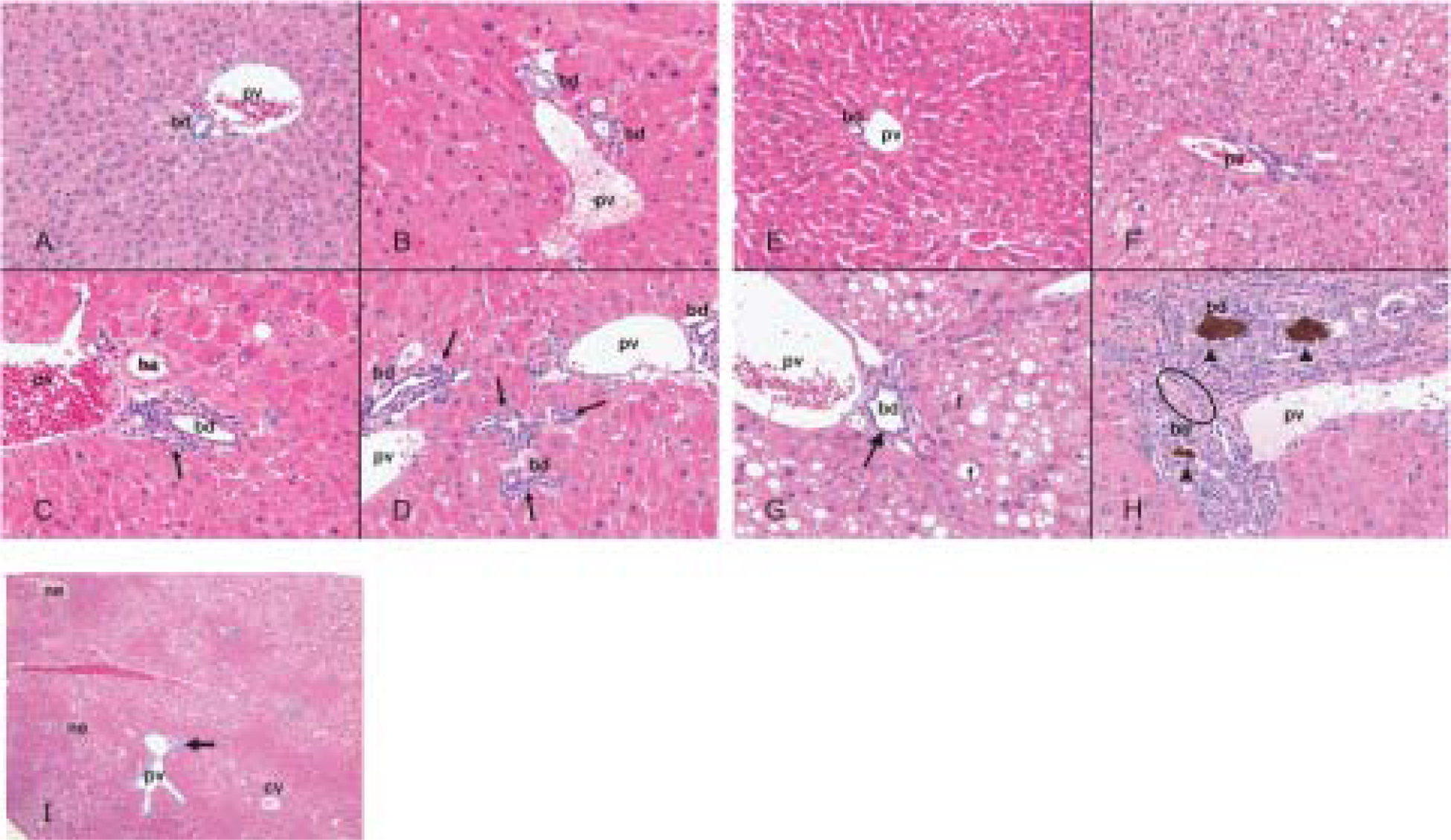
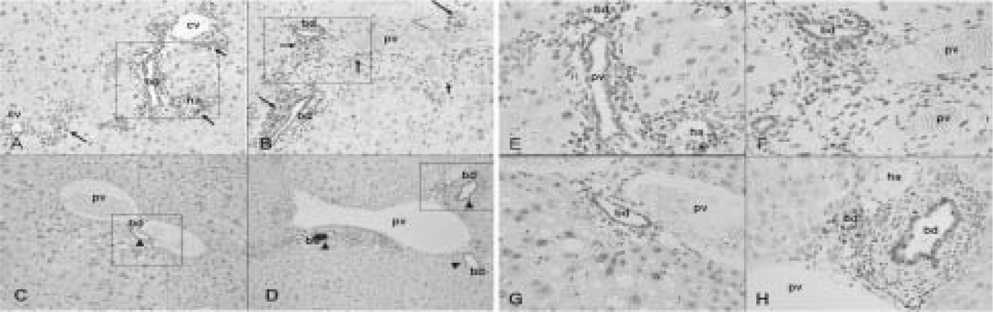
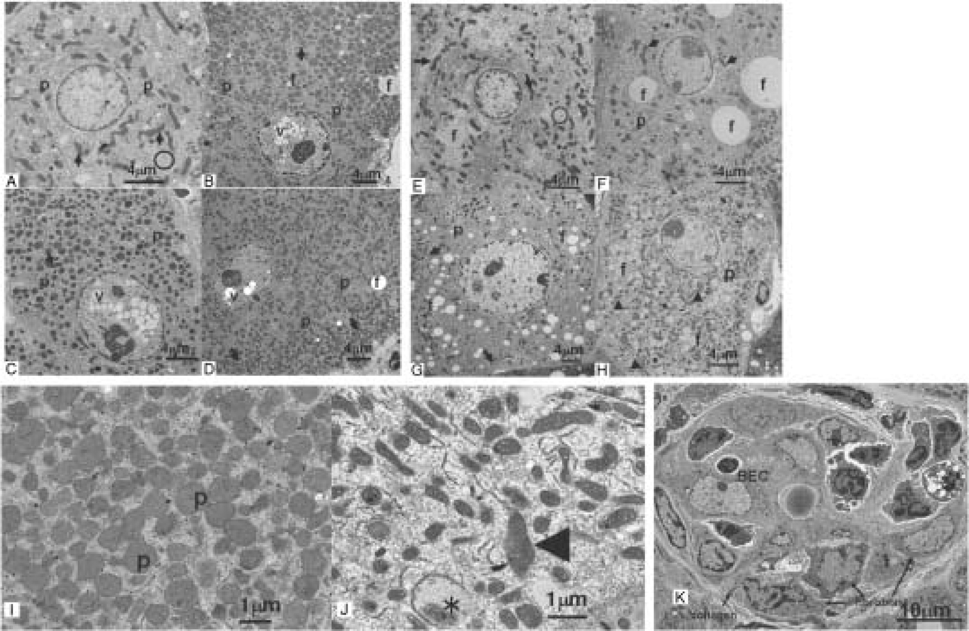
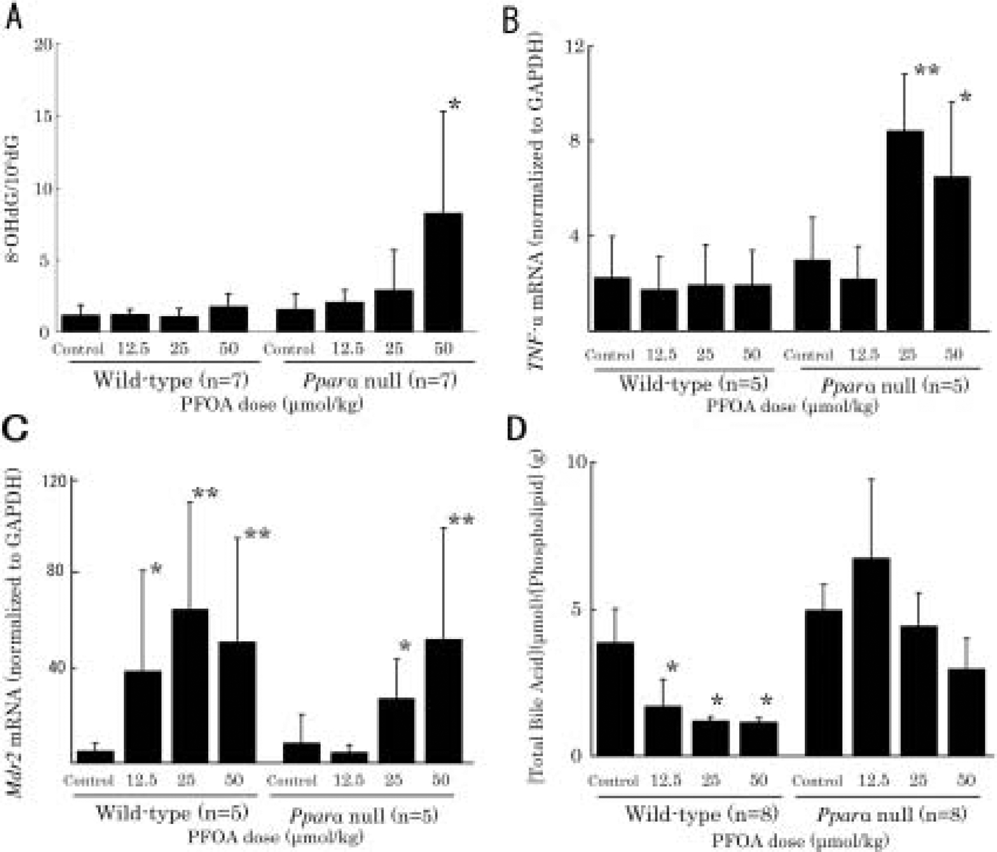
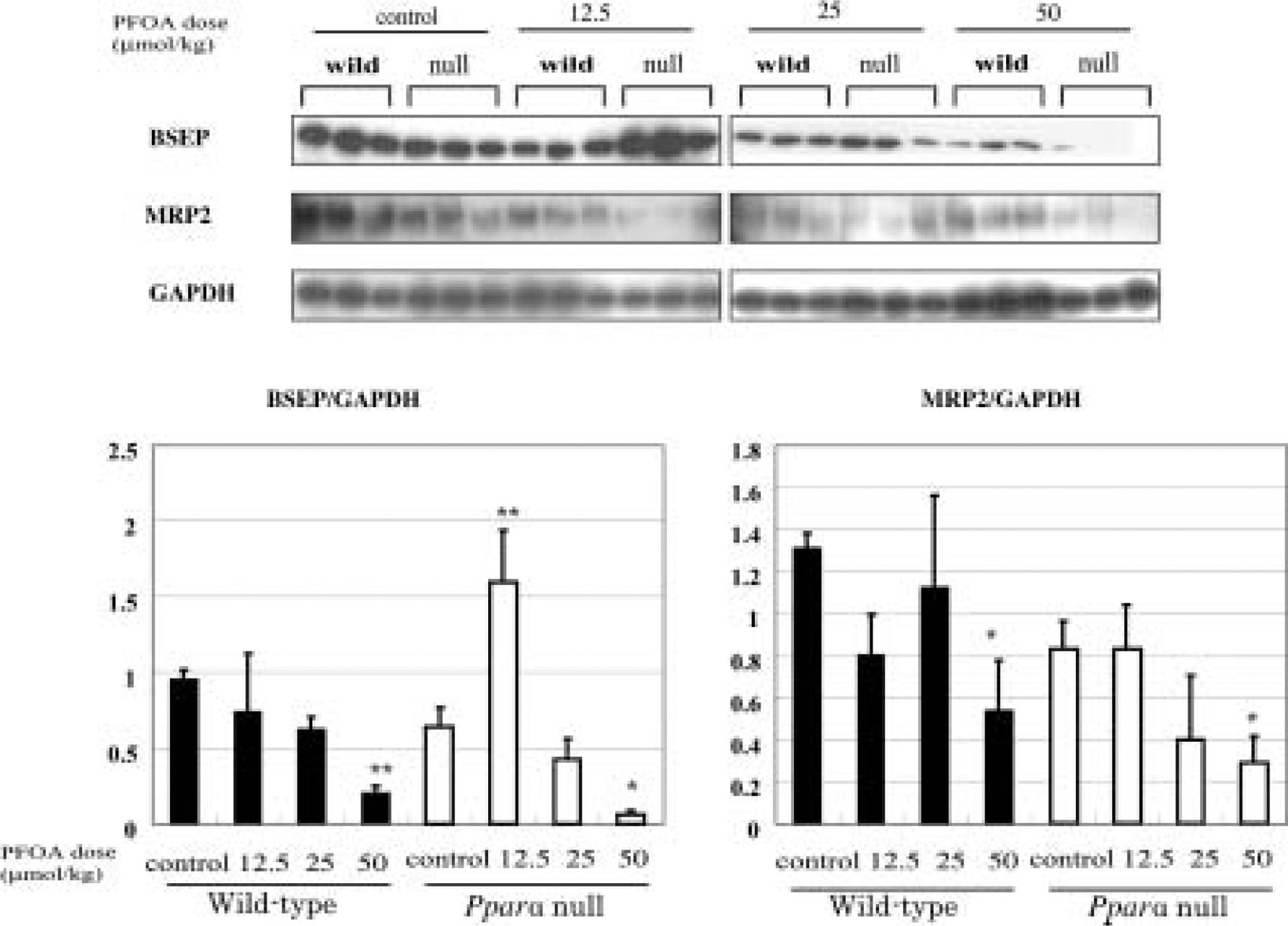
Peroxisome proliferator-activated receptor-alpha (PPARalpha) has been suggested to protect against chemically induced hepatobiliary injuries in rodents. This function could mask the potential toxicities of perfluorooctanoic acid (PFOA) that is an emerging environmental contaminant and a weak ligand of PPARalpha. However its function has not been clarified. In this study, PFOA was found to elicit hepatocyte and bile duct injuries in Pparalpha-null mice after 4 wk treatment with PFOA ammonium salt (0, 12.5, 25, 50 micromol/kg/d, gavage). In wild-type mice, PFOA caused major hepatocellular damage dose-dependently and minor cholangiopathy observed only at 25 and 50 micromol/kg. In treated Pparalpha-null mice, PFOA produced marked fat accumulation, severe cholangiopathy, hepatocellular damage and apoptotic cells especially in bile ducts. Oxidative stress was also increased 4-fold at 50 micromol/kg and TNF-alpha mRNA was upregulated more than 3-fold at 25 micromol/kg in Pparalpha-null mice. Biliary bile acid/phospholipid ratios were higher in Pparalpha-null mice than in wild-type mice. Results from these studies suggest that PPARalpha is protective against PFOA and have a critical role in drug induced hepatobiliary injury.
Figures
Similar articles
-
Modulation of ammonium perfluorooctanoate-induced hepatic damage by genetically different PPARα in mice.Arch Toxicol. 2012 Jan;86(1):63-74. doi: 10.1007/s00204-011-0704-3. Epub 2011 Apr 17. Arch Toxicol. 2012. PMID: 21499893 Free PMC article.
-
Microgram-order ammonium perfluorooctanoate may activate mouse peroxisome proliferator-activated receptor alpha, but not human PPARalpha.Toxicology. 2009 Nov 9;265(1-2):27-33. doi: 10.1016/j.tox.2009.09.004. Epub 2009 Sep 12. Toxicology. 2009. PMID: 19751795 Free PMC article.
-
Gene profiling in the livers of wild-type and PPARalpha-null mice exposed to perfluorooctanoic acid.Toxicol Pathol. 2008 Jun;36(4):592-607. doi: 10.1177/0192623308318208. Epub 2008 May 8. Toxicol Pathol. 2008. PMID: 18467677
-
Perfluorooctanoic Acid (PFOA)-induced Liver Lesions in Two Strains of Mice Following Developmental Exposures: PPARα Is Not Required.Toxicol Pathol. 2015 Jun;43(4):558-68. doi: 10.1177/0192623314558463. Epub 2014 Nov 14. Toxicol Pathol. 2015. PMID: 25398757 Free PMC article.
-
Immunotoxicity of perfluorooctanoic acid and perfluorooctane sulfonate and the role of peroxisome proliferator-activated receptor alpha.Crit Rev Toxicol. 2009;39(1):76-94. doi: 10.1080/10408440802209804. Crit Rev Toxicol. 2009. PMID: 18802816 Review.
Cited by
-
Modulation of ammonium perfluorooctanoate-induced hepatic damage by genetically different PPARα in mice.Arch Toxicol. 2012 Jan;86(1):63-74. doi: 10.1007/s00204-011-0704-3. Epub 2011 Apr 17. Arch Toxicol. 2012. PMID: 21499893 Free PMC article.
-
Exposure to per- and Polyfluoroalkyl Substances and Markers of Liver Injury: A Systematic Review and Meta-Analysis.Environ Health Perspect. 2022 Apr;130(4):46001. doi: 10.1289/EHP10092. Epub 2022 Apr 27. Environ Health Perspect. 2022. PMID: 35475652 Free PMC article.
-
Involvement of oxidative stress and inflammation in liver injury caused by perfluorooctanoic acid exposure in mice.Biomed Res Int. 2014;2014:409837. doi: 10.1155/2014/409837. Epub 2014 Mar 2. Biomed Res Int. 2014. PMID: 24724082 Free PMC article.
-
Associations between per- and polyfluoroalkyl substance exposures and metabolic dysfunction associated steatotic liver disease (MASLD) in adult National Health and Nutrition Examination Survey 2017 to 2018.Toxicol Sci. 2024 Nov 1;202(1):142-151. doi: 10.1093/toxsci/kfae106. Toxicol Sci. 2024. PMID: 39150893 Free PMC article.
-
Celecoxib attenuates hepatosteatosis by impairing de novo lipogenesis via Akt-dependent lipogenic pathway.J Cell Mol Med. 2022 Jul;26(14):3995-4006. doi: 10.1111/jcmm.17435. Epub 2022 Jun 17. J Cell Mol Med. 2022. PMID: 35713152 Free PMC article.
References
-
- Kissa E (2001) Fluorinated surfactants and repellents, 2nd Ed., Marcel Dekker, New York.
-
- Lau C, Anitole K, Hodes C, Lai D, Pfahles-Hutchens A, Seed J (2007) Perfluoroalkyl acids: a review of monitoring and toxicological findings. Toxicol Sci 99, 366–94. - PubMed
-
- Vanden Heuvel JP, Thompson JT, Frame SR, Gillies PJ (2006) Differential activation of nuclear receptors by perfluorinated fatty acid analogs and natural fatty acids: a comparison of human, mouse, and rat peroxisome proliferator-activated receptor-alpha, -beta, and -gamma, liver X receptor-beta, and retinoid X receptor-alpha. Toxicol Sci 92, 476–89. - PubMed
-
- Kennedy GL Jr., Butenhoff JL, Olsen GW, O’Connor JC, Seacat AM, Perkins RG, Biegel LB, Murphy SR, Farrar DG (2004) The toxicology of perfluorooctanoate. Crit Rev Toxicol 34, 351–84. - PubMed