. 2010 Mar 1;9(5):1005-15.
doi: 10.4161/cc.9.5.10936. Epub 2010 Mar 14.

Inhibition of focal adhesion kinase decreases tumor growth in human neuroblastoma

Affiliations

Inhibition of focal adhesion kinase decreases tumor growth in human neuroblastoma

Elizabeth A Beierle et al. Cell Cycle. .

Abstract

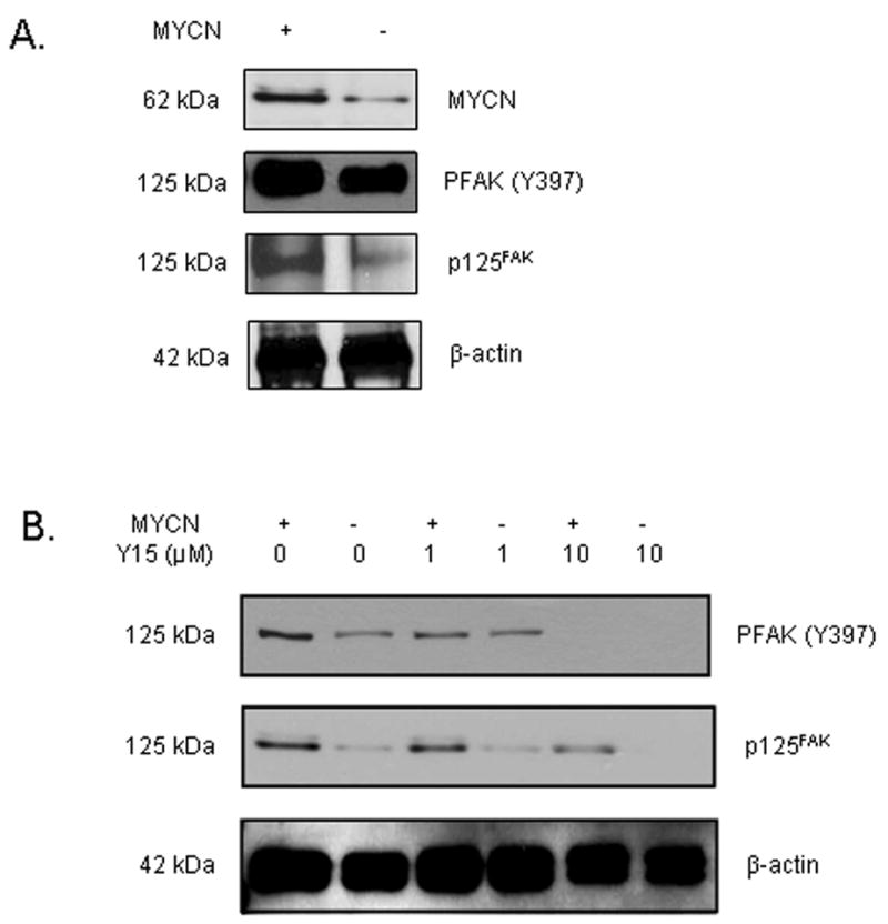
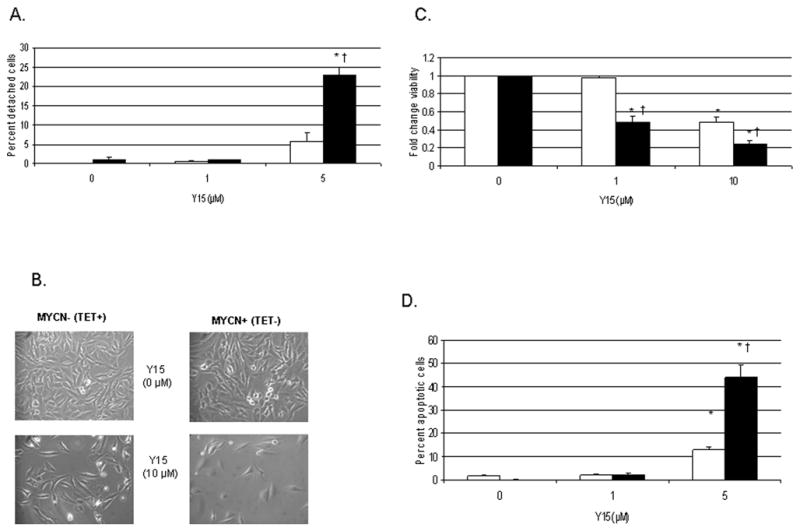
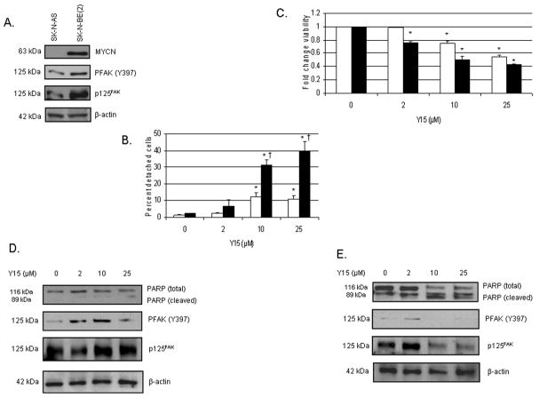
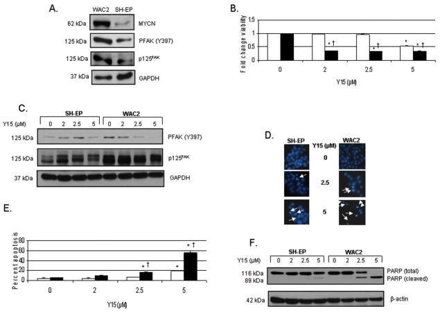
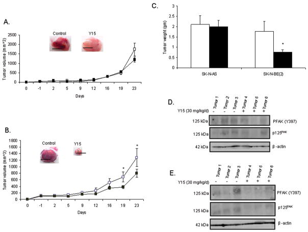
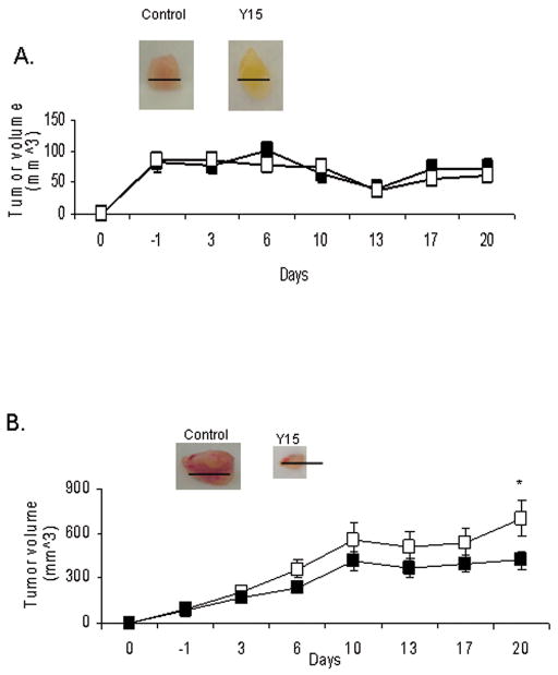
Neuroblastoma is the most common extracranial solid tumor of childhood. Focal adhesion kinase (FAK) is an intracellular kinase that regulates both cellular adhesion and apoptosis. FAK is overexpressed in a number of human tumors including neuroblastoma. Previously, we have shown that the MYCN oncogene, the primary adverse prognostic indicator in neuroblastoma, regulates the expression of FAK in neuroblastoma. In this study, we have examined the effects of FAK inhibition upon neuroblastoma using a small molecule [1,2,4,5-benzenetetraamine tetrahydrochloride (Y15)] to inhibit FAK expression and the phosphorylation of FAK at the Y397 site. Utilizing both non-isogenic and isogenic MYCN(+)/MYCN(-) neuroblastoma cell lines, we found that Y15 effectively diminished phosphorylation of the Y397 site of FAK. Treatment with Y15 resulted in increased detachment, decreased cell viability and increased apoptosis in the neuroblastoma cell lines. We also found that the cell lines with higher MYCN are more sensitive to Y15 treatment than their MYCN negative counterparts. In addition, we have shown that treatment with Y15 in vivo leads to less tumor growth in nude mouse xenograft models, again with the greatest effects seen in MYCN(+) tumor xenografts. The results of the current study suggest that FAK and phosphorylation at the Y397 site plays a role in neuroblastoma cell survival, and that the FAK Y397 phosphorylation site is a potential therapeutic target for this childhood tumor.


Figures

Fig. 1
Fig. 1
The phosphorylation of Y397 FAK is decreased after treatment of MYCN+ (Tet−)/MYCN− (Tet+) isogenic neuroblastoma cell lines with 1,2,4,5-benzenetetraamine tetrahydrochloride (Y15). A, The isogenic MYCN+ (Tet−) and MYCN− (Tet+) neuroblastoma cell lines were examined with immunoblotting. There is increased MYCN, FAK and phosphorylation of Y397 FAK in the MYCN+ (Tet−) compared to the isogenic MYCN− (TET+) cells. B, The isogenic MYCN+ (Tet−) and MYCN− (Tet+) cells were treated with various concentrations of Y15 and cell lysates were examined with immunoblotting. There was a decrease in phosphorylation of Y397 FAK with Y15 treatment. At 1 μM concentration of Y15, there was a greater decrease in phosphorylation of Y397 FAK in the MYCN+ cells. At concentrations of 10 μM, Y15 treatment results in almost complete loss of Y397 phosphorylation in both cell lines.
Fig. 2
Fig. 2
1,2,4,5-benzenetetraamine tetrahydrochloride (Y15) treatment causes cellular detachment, decreased viability and increased apoptosis in human neuroblastoma cell lines. We treated the MYCN+ (Tet−)/MYCN− (Tet+) cells with 1,2,4,5-benzenetetraamine tetrahydrochloride (Y15) to test its effects upon human neuroblastoma cells relative to MYCN status. A, Cellular detachment is measured by counting the number of detached cells and expressing it as a ratio to the total number of cells present, that is detached plus attached cells (mean ± SEM). MYCN− (Tet+) cells are represented by the open bars (□) and MYCN+ (Tet−) cells are represented by the black bars (■). After only 24 hours of treatment with Y15, there is a significant increase in the number of MYCN+ (■) neuroblastoma cells that are detached compared to the MYCN− (□) cells [23 ± 2% vs. 5.6 ± 2.6%; *p≤0.01 control vs. Y15; †p≤0.05 MYCN+ (■) vs. MYCN− (□)]. B, MYCN+ (Tet−) and MYCN− (Tet+) cells are treated with Y15 for 48 hours and cells are examined by phase contrast microscopy. There are significantly fewer cells present after Y15 treatment, with the decrease in cell number being more marked in the MYCN+ (Tet−) cell line (bottom right panel). C, MYCN+ (Tet−) (■) and MYCN− (Tet+) (□) cells are treated with Y15 for 48 hours and viability is measured with MTT assay, expressed as fold change (mean ± SEM). There is a significant decrease in viability in both cell lines compared to non-treated controls, but the decrease is more pronounced in the MYCN+ (■) cell line, which reaches 50% decrease in viability at 1 μM treatment [*p≤0.01 control vs. Y15; †p≤0.05 MYCN+ (■)vs. MYCN− (□)]. D, MYCN+ (Tet−) (■) and MYCN− (Tet+) (□) cells are treated with Y15 for 24 hours and Hoechst staining is used to detect apoptosis, expressed as a percentage of apoptotic cells to non-apoptotic cells (mean ± SEM). Y15 resulted in a significant increase in apoptosis in both cell lines after only 24 hours of treatment, and similar to the results with viability, Y15 had a greater effect upon apoptosis in the MYCN+ (■) cell line compared to the MYCN− (□) cells [44.3 ± 4.8% vs. 13 ± 1.2%, MYCN+ (■) vs. MYCN− (□) at Y15 5 μM; *p≤0.01 control vs. Y15; †p≤0.01 MYCN+ (■) vs. MYCN− (□)].
Fig. 3
Fig. 3
1,2,4,5-benzenetetraamine tetrahydrochloride (Y15) treatment leads to decreased phosphorylation of Y397 FAK, cellular detachment and viability, and increased apoptosis in SK-N-AS and SK-N-BE(2) neuroblastoma cell lines. A, The SK-N-BE(2) (MYCN+) neuroblastoma cells have more total FAK expression and more phosphorylation of Y397 FAK that the SK-N-AS (MYCN−) cell line as shown by immunoblotting. B, Detachment is measured by counting the detached cells and expressing as a ratio of total cells present, that is, detached plus attached cells (mean ± SEM). SK-N-AS (MYCN−) cells are represented by the open bars (□) and SK-N-BE(2) (MYCN+) cells are represented by the black bars (■). Treatment with Y15 leads to cellular detachment in both cell lines, but the SK-N-BE(2) (MYCN+) (■) cells are more sensitive to treatment and have more detachment than the SK-N-AS (MYCN−) (□) cells [*p≤0.01 control vs. Y15; †p≤0.05 SK-N-AS (□) vs. SK-N-BE(2) (■)]. C, MTT assay for cell viability shows that Y15 treatment leads to decreased cell viability, and again, the SK-N-BE(2) (■) cell line is more sensitive to Y15 treatment, reaching a 50% decrease in viability at only 10 μM Y15 compared to the 25 μM required for the SK-N-AS (□) cells to reach 50% viability (*p≤0.01 control vs. Y15). D, Western blotting for evaluation of FAK and apoptosis in the SK-N-AS (MYCN−) cell line reveals some loss of Y397 FAK phosphorylation with Y15 treatment at 25 μM (second panel). In addition, at this highest concentration, there is biochemical evidence of apoptosis with a decrease in total PARP and an increase in cleaved PARP (top panel). E, Western blotting for evaluation of FAK and apoptosis in the SK-N-BE(2) (MYCN+ ) cell line reveals a marked loss of Y397 FAK phosphorylation with Y15 treatment at 10 μM with significant loss of total PARP and increase in cleaved PARP (top panel) at 10 μM as biochemical evidence of apoptosis. These data correspond to the detachment and viability data showing that the SK-N-BE(2) (MYCN+) cell line is more sensitive to inhibition of FAK phosphorylation than the SK-N-AS (MYCN−) cell line.
Fig. 4
Fig. 4
1,2,4,5-benzenetetraamine tetrahydrochloride (Y15) treatment leads to decreased phosphorylation of Y397 FAK, cellular detachment and viability, and increased apoptosis in the isogenic WAC2 (MYCN+) and SH-EP (MYCN−) neuroblastoma cell lines. A, Western blotting confirms that these isogenic neuroblastoma cell lines differ in MYCN, total FAK, and Y397 FAK, with the WAC2 cell line having more MYCN, total FAK and more phosphorylation at the Y397 site of FAK than the SH-EP cell line. B, MTT assay was used to measure cell viability after Y15 treatment. Viability is expressed as mean fold change ± SEM. SH-EP (MYCN−) cells are represented by the open bars (□) and WAC2 (MYCN+) cells are represented by the black bars (■). Treatment of the WAC2 (MYCN+) (■) cell line with Y15 leads to 50% decrease in cell viability at 2 μM concentration. The SH-EP (□) cell line required over twice that concentration of Y15 to reach a 50% decrease in viability [*p≤0.01 control vs. Y15; †p≤0.01 SH-EP (□) vs. WAC2 (■)]. C, Western blotting is utilized to detect FAK and Y397 FAK phosphorylation. Treatment with Y15 leads to loss of Y397 FAK phosphorylation in the WAC2 cell line at 2.5 μM (lane 7) and in the SH-EP cell line at 5 μM (lane 4). D, Hoechst staining is utilized to detect apoptosis. The condensed and fragmented nuclei (white arrows) are easily detected and counted. The loss of Y397 FAK phosphorylation corresponds to increased apoptosis seen in the WAC2 cell line after treatment with Y15 at 2.5 μM (white arrows, middle right panel) and in the SH-EP cells after 5 μM (white arrows, lower left panel). E, Graphical representation of Hoechst staining data. There is a significant increase in the percentage of apoptotic cells in the WAC2 cell line (■) after Y15 treatment at 2.5 μM, and in the SH-EP cells (□) at 5 μM [*p≤0.01 control vs. Y15; †p≤0.01 SH-EP (□) vs. WAC2 (■)]. F, Western blotting to detect total and cleaved PARP shows biochemical corroboration of apoptosis in the SH-EP and WAC2 cell lines consistent with the Hoechst staining data. Again, the MYCN+ WAC2 cells are more sensitive to Y15 treatment than their isogenic MYCN− SH-EP counterparts and show significant changes in cleaved PARP at 2.5 μM Y15 treatment.
Fig. 5
Fig. 5
1,2,4,5-benzenetetraamine tetrahydrochloride (Y15) inhibits human neuroblastoma SK-N-BE(2) neuroblastoma xenografts. A, SK-N-AS tumor cells were injected into the flank of female nude mice, mice were treated daily with control vehicle (normal saline, N=8) or Y15 (30 mg/kg/day, N=12), and tumor volumes were measured. In the Figure, vehicle treated mice are represented by the white squares (□), and Y15 treated mice are represented by the black squares (■). Treatment with Y15 had no significant effect upon SK-N-AS tumor volume. Black bars in the photographs represent 1 cm. B, SK-N-BE(2) tumor cells were injected into the flank of female nude mice, mice were treated daily with control vehicle (normal saline, N=7) or Y15 (30 mg/kg/day, N=13) and tumor volumes were measured. In the Figure, vehicle treated mice are represented by the white squares (□), and Y15 treated mice are represented by the black squares (■). Treatment with Y15 resulted in a significant decrease in tumor volumes in these animals [*p≤0.05 control (□) vs. Y15 (■)]. Black bars in the photograph represent 1cm. C, In the Figure, vehicle treated mice are represented by the white bars (□) and Y15 treated animals by the black bars (■). Tumors were weighed, and there was a significant decrease in tumor weight in the Y15 treated SK-N-BE(2) (MYCN+) (■) neuroblastoma xenografts [*p≤0.01 control (□) vs. Y15 (■)]. Treatment with Y15 did not affect the weights of the SK-N-AS xenografts. D, Tumor xenografts from the SK-N-AS tumors were homogenized and protein was harvested. Western blotting was performed to detect FAK and phosphorylation of Y397 FAK. Y15 treatment results in little change in phosphorylation of Y397 or FAK in the SK-N-AS (MYCN−) tumor xenografts. E, Tumor xenografts from the SK-N-BE(2) tumors were homogenized and protein was harvested. Western blotting was performed to detect FAK and phosphorylation of Y397 FAK. In contrast to the SK-N-AS (MYCN−) tumor xenografts, in the SK-N-BE(2) (MYCN+) tumor xenografts, Y15 treatment results in a decrease in FAK and in Y397 FAK phosphorylation.
Fig. 6
Fig. 6
1,2,4,5-benzenetetraamine tetrahydrochloride (Y15) inhibits human neuroblastoma WAC2 (MYCN+) neuroblastoma xenografts, but has no effect upon SH-EP (MYCN−) xenografts. A, SH-EP (MYCN−) human neuroblastoma tumor cells were injected into the flank of female nude mice, mice were treated daily with control vehicle (normal saline, N=11) or Y15 (30 mg/kg/day, N=15), and tumor volumes were measured. Vehicle treated mice are represented by the white squares (□), and Y15 treated mice are represented by the black squares (■). Treatment with Y15 had no significant effect upon SH-EP tumor volume. Black bars in the Figure represent 100 mm. B, WAC2 (MYCN+) human neuroblastoma tumor cells were injected into the flank of female nude mice, the mice were treated with control vehicle (normal saline, N=10) or Y15 (30 mg/kg/day, N=15) daily, and tumor volumes were measured. In the Figure, vehicle treated mice are represented by the white squares (□), and Y15 treated mice are represented by the black squares (■). Treatment of the WAC2 tumor xenografts with Y15 resulted in a significant decrease in tumor volume [*p≤0.01 control (□) vs. Y15 (■)]. Black bars in the Figure represent 1cm.

Comment in

References

    1. Maris JM, Matthay KK. Molecular biology of neuroblastoma. J Clin Oncol. 1999;17(7):2263–79. - PubMed
    1. Cotterill SJ, Parker L, More L, Craft AW. Neuroblastoma: changing incidence and survival in young people aged 0–24 years. A report from the North of England Young Persons’ Malignant Disease Registry. Med Pediatr Oncol. 2001;36(1):231–4. - PubMed
    1. Brodeur GM, Seeger RC, Schwab M, Varmus HE, Bishop JM. Amplification of N-myc in untreated human neuroblastomas correlates with advanced stage disease. Science. 1984;224(4653):1121–4. - PubMed
    1. Seeger RC, Brodeur GM, Sather H, Dalton A, Siegle SE, Wong KY, et al. Association of multiple copies of the N-myc oncogene with rapid progression of neuroblastomas. N Engl J Med. 1985;313(18):1111–6. - PubMed
    1. Zaizen Y, Taniguchi S, Noguchi S, Suita S. The effect of N-myc amplification and expression on invasiveness of neuroblastoma cells. J Pediatr Surg. 1993;28(6):766–9. - PubMed

Publication types

MeSH terms