Epstein-Barr virus in Burkitt's lymphoma: a role for latent membrane protein 2A
- PMID: 20160479
- PMCID: PMC2855765
- DOI: 10.4161/cc.9.5.10840
Epstein-Barr virus in Burkitt's lymphoma: a role for latent membrane protein 2A
Abstract
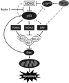
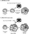
Burkitt's lymphoma (BL) is characterized by translocation of the MYC gene to an immunoglobulin locus. Transgenic mouse models have been used to study the molecular changes that are necessary to bypass tumor suppression in the presence of translocated MYC. Inactivation of the p53 pathway is a major step to tumor formation in mouse models that is also seen in human disease. Human BL is often highly associated with Epstein-Barr virus (EBV). The EBV latency protein latent membrane protein 2A (LMP2A) is known to promote B cell survival by affecting levels of pro-survival factors. Using LMP2A transgenic mouse models, we have identified a novel mechanism that permits lymphomagenesis in the presence of an intact p53 pathway. This work uncovers a contribution of EBV to molecular events that have documented importance in BL pathogenesis, and may underlie the poorly understood link between EBV and BL.
Figures


Similar articles
-
Latent membrane proteins from EBV differentially target cellular pathways to accelerate MYC-induced lymphomagenesis.Blood Adv. 2022 Jul 26;6(14):4283-4296. doi: 10.1182/bloodadvances.2022007695. Blood Adv. 2022. PMID: 35605249 Free PMC article.
-
Rapamycin reverses splenomegaly and inhibits tumor development in a transgenic model of Epstein-Barr virus-related Burkitt's lymphoma.Mol Cancer Ther. 2011 Apr;10(4):679-86. doi: 10.1158/1535-7163.MCT-10-0833. Epub 2011 Jan 31. Mol Cancer Ther. 2011. PMID: 21282357 Free PMC article.
-
Epstein-Barr virus LMP2A bypasses p53 inactivation in a MYC model of lymphomagenesis.Proc Natl Acad Sci U S A. 2009 Oct 20;106(42):17945-50. doi: 10.1073/pnas.0907994106. Epub 2009 Oct 7. Proc Natl Acad Sci U S A. 2009. PMID: 19815507 Free PMC article.
-
How does Epstein-Barr virus (EBV) complement the activation of Myc in the pathogenesis of Burkitt's lymphoma?Semin Cancer Biol. 2009 Dec;19(6):366-76. doi: 10.1016/j.semcancer.2009.07.007. Epub 2009 Jul 25. Semin Cancer Biol. 2009. PMID: 19635566 Free PMC article. Review.
-
The signaling pathways of Epstein-Barr virus-encoded latent membrane protein 2A (LMP2A) in latency and cancer.Cell Mol Biol Lett. 2009;14(2):222-47. doi: 10.2478/s11658-008-0045-2. Epub 2008 Dec 13. Cell Mol Biol Lett. 2009. PMID: 19082921 Free PMC article. Review.
Cited by
-
EBV and Apoptosis: The Viral Master Regulator of Cell Fate?Viruses. 2017 Nov 13;9(11):339. doi: 10.3390/v9110339. Viruses. 2017. PMID: 29137176 Free PMC article. Review.
-
The impact of Epstein-Barr virus latent membrane protein 2A on the production of B cell activating factor of the tumor necrosis factor family (BAFF), APRIL and their receptors.Immun Inflamm Dis. 2022 Nov;10(11):e729. doi: 10.1002/iid3.729. Immun Inflamm Dis. 2022. PMID: 36301035 Free PMC article.
-
The role of EBV in the pathogenesis of Burkitt's Lymphoma: an Italian hospital based survey.Infect Agent Cancer. 2014 Oct 15;9(1):34. doi: 10.1186/1750-9378-9-34. eCollection 2014. Infect Agent Cancer. 2014. PMID: 25364378 Free PMC article. Review.
-
Identification of protein kinase inhibitors with a selective negative effect on the viability of Epstein-Barr virus infected B cell lines.PLoS One. 2014 Apr 23;9(4):e95688. doi: 10.1371/journal.pone.0095688. eCollection 2014. PLoS One. 2014. PMID: 24759913 Free PMC article.
-
A shared gene expression signature in mouse models of EBV-associated and non-EBV-associated Burkitt lymphoma.Blood. 2011 Dec 22;118(26):6849-59. doi: 10.1182/blood-2011-02-338434. Epub 2011 Oct 28. Blood. 2011. PMID: 22039254 Free PMC article.
References
-
- Burkitt D. A sarcoma involving the jaws in African children. Br J Surg. 1958;46:218–223. - PubMed
-
- Burkitt D, O'Conor GT. Malignant lymphoma in African children. I. A clinical syndrome. Cancer. 1961;14:258–269. - PubMed
-
- Epstein MA. The Origins of EBV Research. In: Robertson ES, editor. Epstein-Barr Virus. Norfolk, England: Caister Academic Press; 2005. pp. 1–14.
-
- Epstein MA, Barr YM. Cultivation in Vitro of Human Lymphoblasts from Burkitt's Malignant Lymphoma. Lancet. 1964;1:252–253. - PubMed
-
- Epstein MA, Achong BG, Barr YM. Virus Particles in Cultured Lymphoblasts from Burkitt's Lymphoma. Lancet. 1964;1:702–703. - PubMed
Publication types
MeSH terms
Substances
Grants and funding
- T32 CA009560/CA/NCI NIH HHS/United States
- CA117794/CA/NCI NIH HHS/United States
- AI067048/AI/NIAID NIH HHS/United States
- R01 CA073507/CA/NCI NIH HHS/United States
- CA73507/CA/NCI NIH HHS/United States
- CA133063/CA/NCI NIH HHS/United States
- AI076183/AI/NIAID NIH HHS/United States
- R01 CA117794/CA/NCI NIH HHS/United States
- T32CA009560/CA/NCI NIH HHS/United States
- R01 CA133063/CA/NCI NIH HHS/United States
- R01 AI067048/AI/NIAID NIH HHS/United States
- CA021776/CA/NCI NIH HHS/United States
- R01 CA021776/CA/NCI NIH HHS/United States
- R01 AI076183/AI/NIAID NIH HHS/United States
LinkOut - more resources
Full Text Sources
Research Materials
Miscellaneous
