. 2009 Oct 13;5(10):2909-2923.
doi: 10.1021/ct900262t.

An Evaluation of Explicit Receptor Flexibility in Molecular Docking Using Molecular Dynamics and Torsion Angle Molecular Dynamics

Affiliations

An Evaluation of Explicit Receptor Flexibility in Molecular Docking Using Molecular Dynamics and Torsion Angle Molecular Dynamics

Roger S Armen et al. J Chem Theory Comput. .

Abstract

Incorporating receptor flexibility into molecular docking should improve results for flexible proteins. However, the incorporation of explicit all-atom flexibility with molecular dynamics for the entire protein chain may also introduce significant error and "noise" that could decrease docking accuracy and deteriorate the ability of a scoring function to rank native-like poses. We address this apparent paradox by comparing the success of several flexible receptor models in cross-docking and multiple receptor ensemble docking for p38α mitogen-activated protein (MAP) kinase. Explicit all-atom receptor flexibility has been incorporated into a CHARMM-based molecular docking method (CDOCKER) using both molecular dynamics (MD) and torsion angle molecular dynamics (TAMD) for the refinement of predicted protein-ligand binding geometries. These flexible receptor models have been evaluated, and the accuracy and efficiency of TAMD sampling is directly compared to MD sampling. Several flexible receptor models are compared, encompassing flexible side chains, flexible loops, multiple flexible backbone segments, and treatment of the entire chain as flexible. We find that although including side chain and some backbone flexibility is required for improved docking accuracy as expected, docking accuracy also diminishes as additional and unnecessary receptor flexibility is included into the conformational search space. Ensemble docking results demonstrate that including protein flexibility leads to to improved agreement with binding data for 227 active compounds. This comparison also demonstrates that a flexible receptor model enriches high affinity compound identification without significantly increasing the number of false positives from low affinity compounds.


Figures
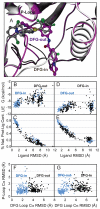
Figure 1
Figure 1
A successful cross docking for a difficult example involving DFG loop rearrangement. (A) the superposition of a DFG-in protein-ligand complex (1ouy shown in gray), and a DFG-out protein-ligand complex (1w83 shown in magenta). (B) Ligand RMSD vs. LIE1 score plot of flexible receptor self-docking 1ouy (light blue), compared to flexible receptor cross docking (black) of the ligand 1ouy into the receptor 1w83. (C) Ligand RMSD vs. % native protein-ligand contacts for the same. (D) Ligand RMSD vs. LIE1 score plot of flexible receptor self-docking 1w83 (light blue), compared to flexible receptor cross docking (black) of the ligand 1w83 into the receptor 1ouy. (E) Ligand RMSD vs. % native protein-ligand contacts for the same. (F) Cα RMSD of the P-loop vs. Cα RMSD of the DFG loop plot for flexible receptor self-docking 1ouy (light blue), compared to flexible receptor cross-docking (black) of the ligand 1ouy into the receptor 1w83. (G) Cα RMSD of the P-loop vs. Cα RMSD of the DFG loop plot for flexible receptor self-docking 1w83 (light blue), compared to flexible receptor cross-docking (black) of the ligand 1w83 into the receptor 1ouy.
Figure 2
Figure 2
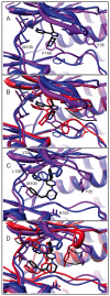
Successful cross-docking examples involving concerted rearrangements of backbone and side chain of the P-loop and the DFG loop. For all images, the reference receptor conformation is shown in blue, superimposed with the starting receptor conformation for a difficult cross-dock shown in purple. The reference ligand position is shown in black. In (B and D), the new lowest energy flexible receptor conformation is shown in red, along with the correctly predicted native-like ligand conformation (RMSD < 2.0 Å). In both examples, Tyr 35 from the P-loop undergoes a successful rearrangement from the starting conformation (purple) to the final conformation (red), which is in turn closer to the reference target conformation (blue) allowing the ligand (red) to dock in the correct conformation. Arrows are shown to highlight the movement of Tyr 35 from the starting conformation closer to the reference conformation. In (A and B), the ligand 1bl7 (DFG-in) is able to dock accurately into the DFG-out conformation because of the backbone and side chain rearrangement of Phe 169. In the second example (C and D) the ligand 1yqj (DFG-in) is able to dock accurately into the 1di9 (DFG-in) receptor conformation.
Figure 3
Figure 3
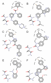
Two common protein-ligand interactions (anchor points), shared by all six series of active compounds binding in the DFG-in receptor conformation. An interaction with the hydrophobic pocket (dashed line) and hydrogen bonding to residues in the gatekeeper region are common to all six series of active compounds: (A) 2-aminopyrimidine carbamates, (B) 4-azaindoles, (C) aminopyrazoles, (D) benzimidazolones, (E) indole and pyridine fragments, and (F) triazolopyridine oxazoles.
Figure 4
Figure 4
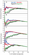
Enrichment factors for rigid and flexible receptor docking into receptors (A) 1a9u, (B) 1bl6, (C) 1kv2, and (D) 1w84. These enrichment factors EF1 and EF2 are calculated assuming that groups of high affinity compounds can be separated from groups of low affinity compounds.
Figure 5
Figure 5
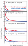
Enrichment factors for separating active compounds from more than 9000 decoy molecules. Enrichment of all 227 actives from decoys for receptor (A) 1a9u and (B) 1kv2. Enrichment of 153 actives in the same molecular weight range (320 to 450) as the decoys for receptor (C) 1a9u and (D) 1kv2.
Figure 6
Figure 6
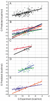
Comparison to binding data, using a minimal ensemble of 4 rigid receptor conformations (1a9u, 1di9, 1ouk, 1oz1). The best linear fit for this model through all points is shown in (A) in black. The best linear fit is shown in (B, C, D, and E) for each of the six series: 1. 2-aminopyrimidine carbamates (red), 2. 4-azaindoles (black), 3. aminopyrazoles (light blue), 4. benzimidazolones (green), 5. indole and pyridine fragments (orange), and 6. triazolopyridine oxazoles (purple).
Figure 7
Figure 7
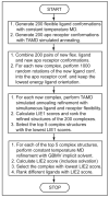
Overview of the flexible receptor docking protocol.

Similar articles

Cited by

References

    1. Carlson HA. Curr. Opin. Chem. Biol. 2002;6:447–52. - PubMed
    1. Abagyan R, Totrov M. Curr. Opin. Chem. Biol. 2001;5:375–82. - PubMed
    1. Gerstein M, Krebs W. Nucleic Acids Res. 1998;26:4280–90. - PMC - PubMed
    1. Kuntz ID, Meng EC, Shoichet BK. Acc. Chem. Res. 1994;27:117–123.
    1. Polgar T, Baki A, Szendrei GI, Keseru GM. J. Med. Chem. 2005;48:7946–59. - PubMed

LinkOut - more resources