

doi: 10.1016/j.jmmm.2009.02.066.
Formation and properties of magnetic chains for 100 nm nanoparticles used in separations of molecules and cells
Affiliations
- PMID: 20161001
- PMCID: PMC2757286
- DOI: 10.1016/j.jmmm.2009.02.066
Item in Clipboard
Formation and properties of magnetic chains for 100 nm nanoparticles used in separations of molecules and cells
J Magn Magn Mater.
.
Abstract
Optical observations of 100 nm metallic magnetic nanoparticles are used to study their magnetic field induced self assembly. Chains with lengths of tens of microns are observed to form within minutes at nanoparticle concentrations of 10(10) per mL. Chain rotation and magnetophoresis are readily observed, and SEM reveals that long chains are not simple single particle filaments. Similar chains are detected for several 100 nm commercial bio-separation nanoparticles. We demonstrate the staged magnetic condensation of different types of nanoparticles into composite structures and show that magnetic chains bind to immunomagnetically labeled cells, serving as temporary handles which allow novel magnetic cell manipulations.
Figures
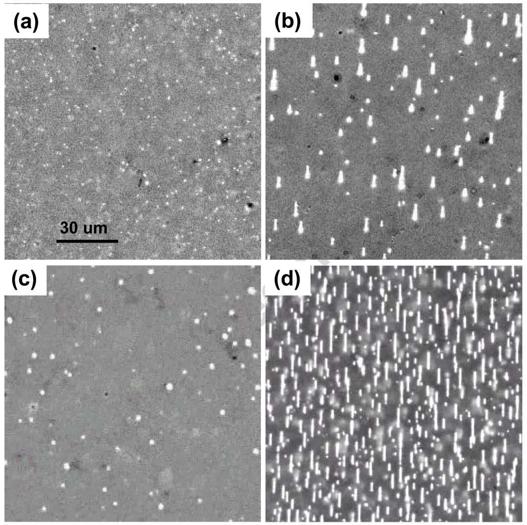
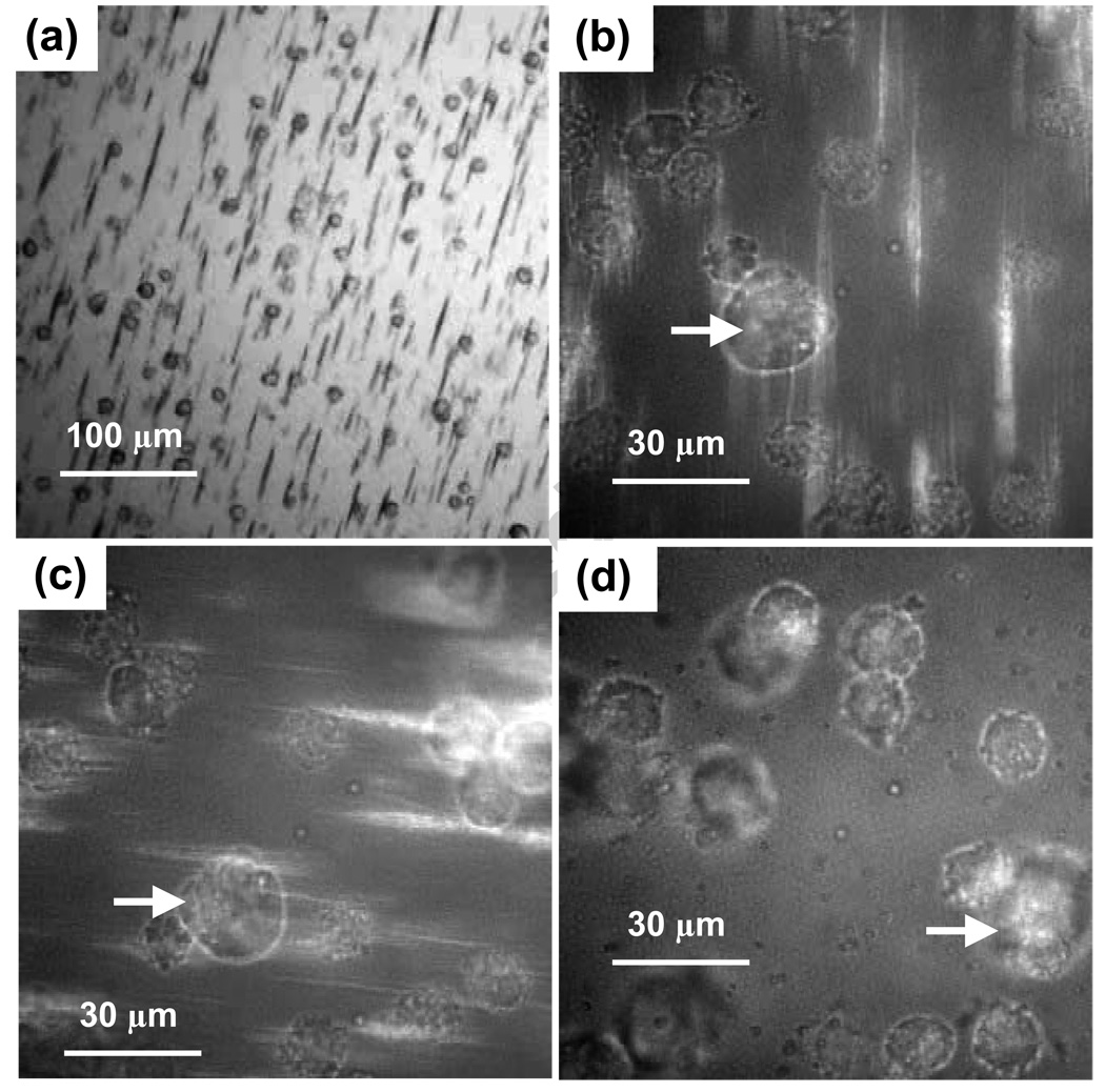
(a) Images of SAF nanoparticles in a 20 µm deep chamber slide in zero field and at 1010/ mL initial concentration. (b) Chain structures formed 50 seconds after H was increased to 1 kOe, 1010/ mL. (c) Small chains which form under the same conditions, except the concentration is 109/ mL. (d) Numerous chains formed in 15 sec when the concentration is 1011/ mL.
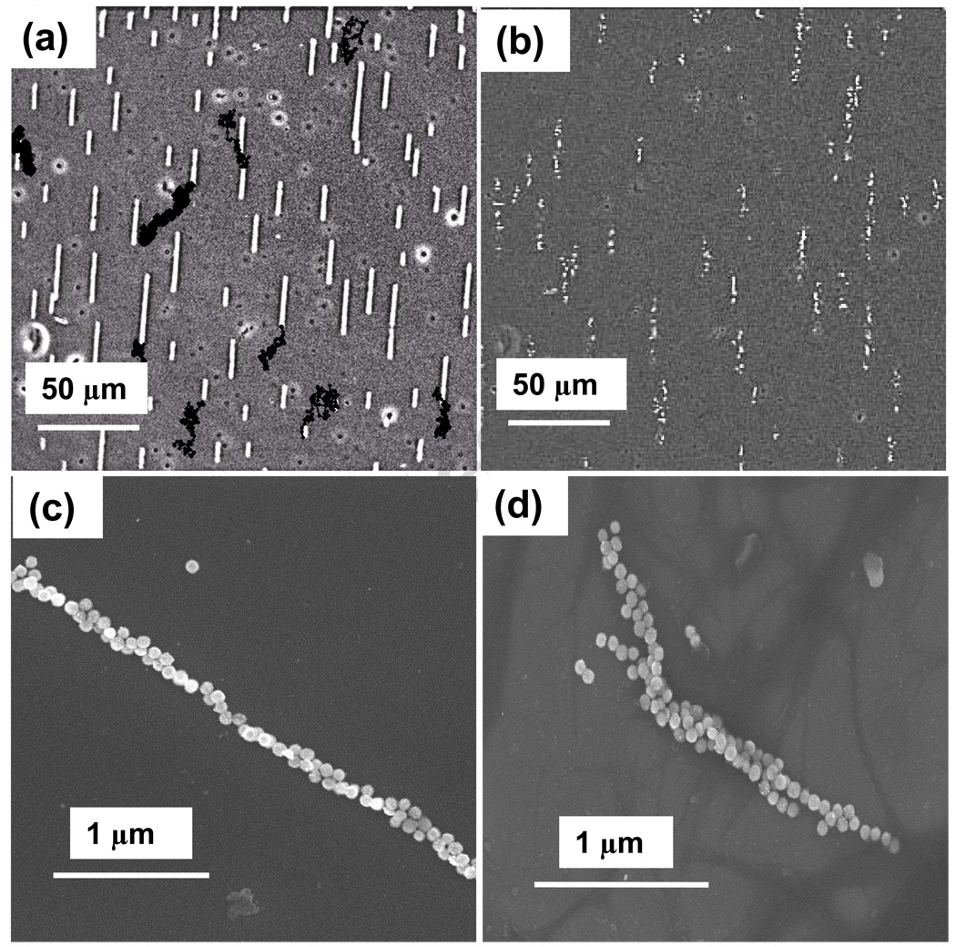
(a) Image showing chains formed with 1010/ mL after 10 minutes. Black dots show selected chain endpoint positions at 10 sec intervals, indicating substantial chain diffusion. (b) The chains in (a) begin disintegrating seconds after the applied field is removed. (c) SEMs reveal that chains are not ideal single particle filaments, and that some chains appear to be composites formed from several chains.
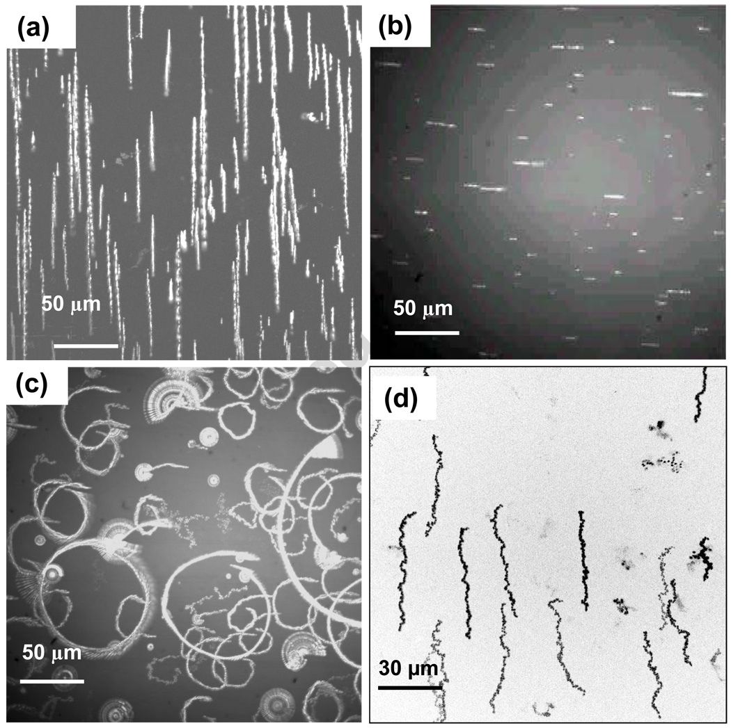
(a) Chain trajectories during gradient driven magnetophoresis are rendered using digital multiple exposures. (b) Initial view of chains in an aged sample, where the magnet is rotated to create circular magnetophoretic trajectories shown in (c). Some chains are susceptible to rotations about fixed surface binding points, but are not free to translate. (d) Vertical orientation of chains in thin cells alters optical contrast, making vertical chains appear dark, and limits chain length and mobility heterogeneities.
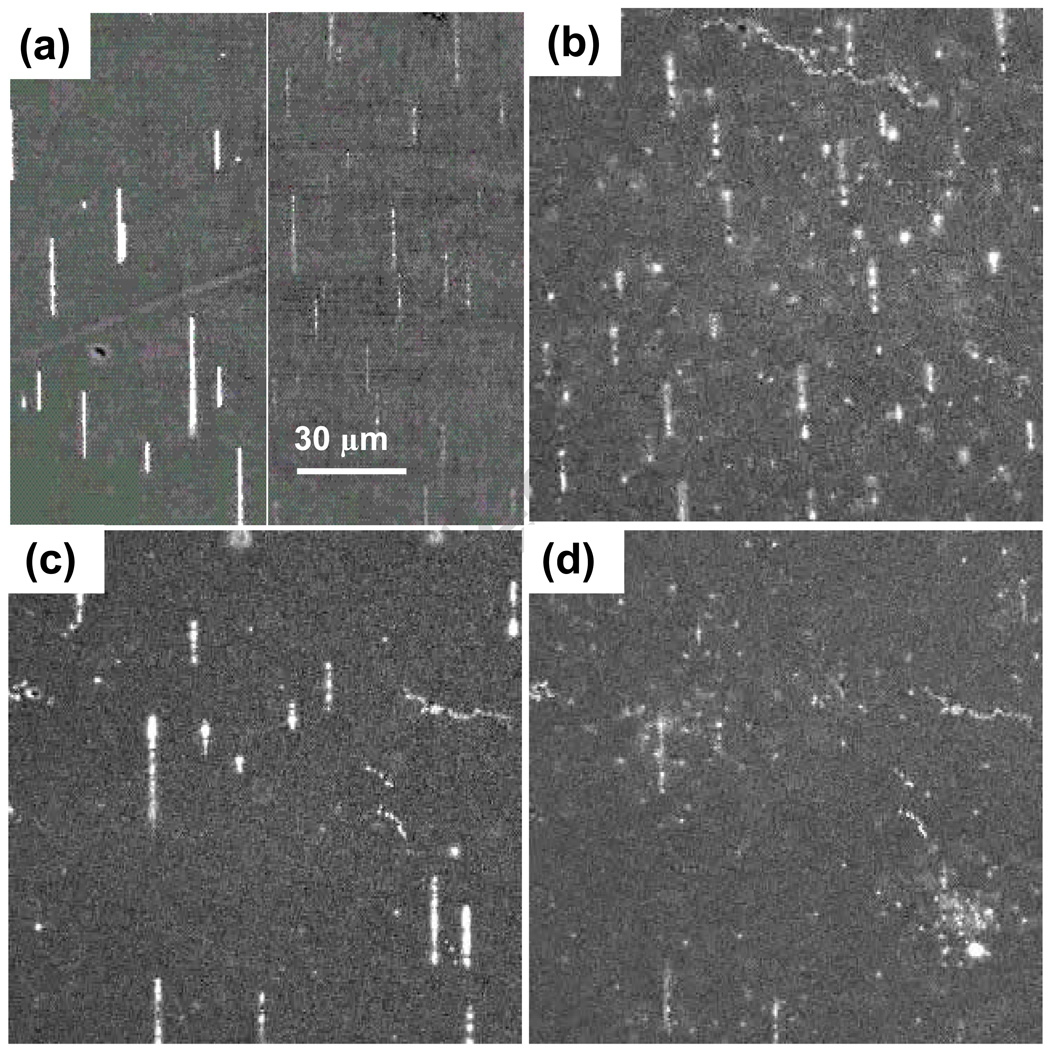
(a) Composite image showing bright contrast of metallic nanoparticle chains (left) and fainter chains of MagCellect chains (right) in the same cell. (b) At 100 Oe, the MC particles chain while the high saturation field metal nanoparticles diffuse rapidly. (c) When the field is increased, SAF particles attach to the chains, brightening them and depleting bright isolated nanoparticles. (d) When the field is reduced to 100 Oe, the metal particles detach.
(a) An image showing MagCellect chains which form on a slide surface that is covered with settled immuno-magnetically labeled cells. (b) A higher magnification view shows cells interacting with chains. (c) Rotation of the magnetic field in the slide plane leads to corresponding rotations for cells attached to chains. (d) Out of plane rotations produce an end-over-end motion of chain-cell assemblies, resulting in lateral translations. Arrows mark the same cell in different frames.
References
-
- Zborowski M, Chalmers JJ, editors. Magnetic Cell Separation, in Laboratory techniques in biochemistry and molecular biology. Vol. 32 2007.
-
- Perez JM, Josephson L, Weissleder R. Chem. BioChem. 2004;5:261. - PubMed
-
- Li GX, Joshi V, White RL, et al. J. Appl. Phys. 2003;93:7557.
-
- Kettering M, Winter J, Zeisberger M, et al. Nanotechnol. 2007;18:175101.
-
- Muthana M, Scott SD, Farrow N, et al. Gene Therapy. 2008;15:902. - PubMed
Grants and funding
LinkOut - more resources
Full Text Sources
Other Literature Sources