. 2009 Oct 5;4(5):414-428.
doi: 10.1016/j.nantod.2009.07.004.

Multifunctional Quantum Dots for Personalized Medicine

Affiliations

Multifunctional Quantum Dots for Personalized Medicine

Pavel Zrazhevskiy et al. Nano Today. .

Abstract

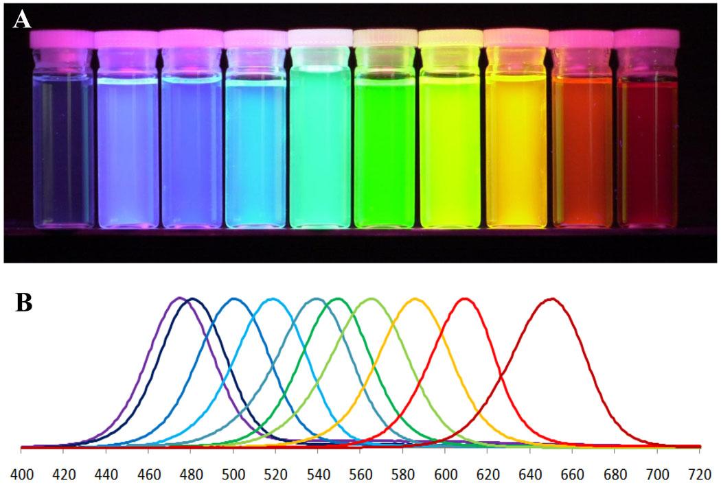
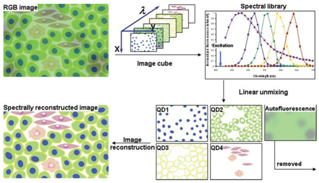
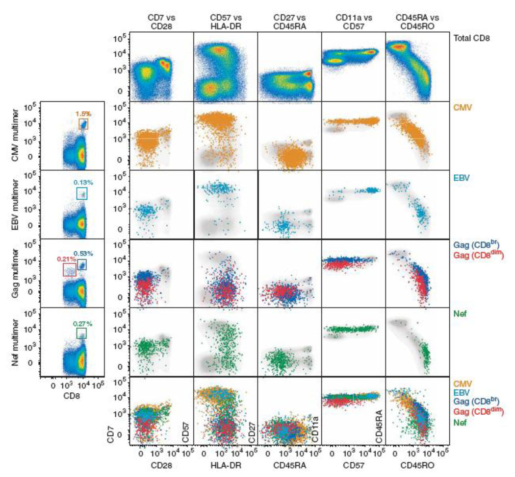
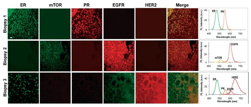
Successes in biomedical research and state-of-the-art medicine have undoubtedly improved the quality of life. However, a number of diseases, such as cancer, immunodeficiencies, and neurological disorders, still evade conventional diagnostic and therapeutic approaches. A transformation towards personalized medicine may help to combat these diseases. For this, identification of disease molecular fingerprints and their association with prognosis and targeted therapy must become available. Quantum dots (QDs), semiconductor nanocrystals with unique photo-physical properties, represent a novel class of fluorescence probes to address many of the needs of personalized medicine. This review outlines the properties of QDs that make them a suitable platform for advancing personalized medicine, examines several proof-of-concept studies showing utility of QDs for clinically relevant applications, and discusses current challenges in introducing QDs into clinical practice.


Figures

Figure 1
Figure 1
Quantum dots possess unique photo-physical properties suitable for addressing the needs of personalized medicine. The ability to utilize multicolor QD probes (A) and tune the emission color by the particle size allows multiplexed biomarker detection. Narrow emission spectra (B) along with efficient light absorption throughout a wide spectrum enable simultaneous imaging of several biomarkers critical for molecular profiling of diseases.
Figure 2
Figure 2
Hyperspectral imaging represents a powerful technique for analysis of multiple QD-labeled biomarkers within a single specimen. While standard RGB camera cannot distinguish spectrally overlapping probes and is limited to analysis of few biomarkers, hyperspectral imaging applies narrow band-pass filters and takes a series of images for each wavelength, thus providing spectral information for each pixel of an image. Further application of spectral library allows accurate unmixing of individual spectrally-distinct components, enhancing the ability for molecular profiling [3].
Figure 3
Figure 3
Seventeen-parameter flow-cytometry analysis of antigen-specific T-cell populations was achieved using 8 QD probes and 9 organic fluorophores. Significant heterogeneity in biomarker expression within a CD8+ T-cell population (shown in gray) emphasizes the need for multi-parameter analysis in studying immune response and other complex systems [25].
Figure 4
Figure 4
Five-parameter quantitative analysis of the three tissue specimens obtained from tumor biopsies clearly identified the differences in biomarker expression profiles between different types of breast cancers. Molecular fingerprinting might not only provide more accurate diagnosis and prognosis, but also identify suitable molecular targets for anti-cancer therapy [23].
Figure 5
Figure 5
Multi-parameter FISH using QD probes and organic fluorophores enables high-resolution imaging of different mRNA molecular within single cells, thus providing information about relative gene expression levels, localization of mRNA within cellular compartments, and co-localization of different mRNA molecules and other biomarkers [28].
Figure 6
Figure 6
Outstanding photostability and high brightness of QD probes enable long-term real-time monitoring of erbB receptor activation by QD-EGF and study the retrograde transport of these probes along the filopodia towards the cell body. Scale bars 5 um [32].
Figure 7
Figure 7
Delivery of QD probes inside cells represents a challenge for labeling intracellular targets. Different modes of delivery are being developed to overcome this issue. A) Mechano-chemical modes of QD delivery involve utilization of mechanically strong materials capable of puncturing cell membrane and reaching into intracellular compartments. Delivery using nanoneedle (left) involves attachment of QDs on the outer surface of a stiff nanotube and manual manipulation of the needle on the cell-by-cell basis [37], while delivery platform based on nanosyringes (right) utilizes arrays of hollow vertically aligned nanotubes that enable intracellular release of QD probes upon cell growth on top of these arrays [38]. B) Encapsulation of QD probes within materials capable of endosomal escape represents a promising high-throughput technique for intracellular QD delivery. A general approach involves coating of QDs with materials possessing proton sponge functionality or other means of destabilizing endosome membrane and functionalizing the surface with targeting ligand. Once ligand binds to a receptor on the cell surface, nanoparticles are uptaken by endocytosis. Decrease in pH inside the endosome causes physical changes in QD coating (usually in surface charge), which triggers mechanisms for cytosolic release of QDs and enables targeting of intracellular components [40].
Figure 8
Figure 8
Utilization of large Stokes shift produced by red and NIR QD probes enables targeted in vivo imaging of subcutaneous tumors. Further image processing with spectral demixing allows efficient removal of tissue autofluorescence [49].
Figure 9
Figure 9
Shallow depth of in vivo imaging with QD probes imposes significant limits on utilization of this technology for deep-tissue imaging. One way to improve the depth and sensitivity of imaging is to use self-illuminating QDs. QD probes conjugated with luciferase convert bioluminescense produced by the enzyme into QD fluorescence emitted in NIR region, thus eliminating autofluorescence and signal intensity attenuation by tissues [55].
Figure 10
Figure 10
Multi-photon microscopy represents a powerful tool for multiplexed in vivo imaging. By utilizing low-energy photons (minimally absorbed by tissues) for excitation of multicolor QD probes, this technique provides deeper tissue penetration and higher sensitivity of imaging. Application of this technique enabled the study of tumor morphology using QDs for labeling of tumor vasculature (blue QDs in (A) and red QDs in (B)), further enhanced by GFP labeling of perivascular cells (green in (A)) and detection of second harmonic generation signal from collagen to visualize extracellular matrix (light-blue in (B)) [58].
Figure 11
Figure 11
QD-based drug carriers integrate drug delivery tracing, loading of various types of drugs (e.g. hydrophobic small-molecule drugs between the QD core and polymer coating or hydrophilic drugs on the exterior surface of the polymeric shell), and targeting functionality. Intermediate size of such carriers ensures slower renal filtration as well as RES uptake, thus increasing blood circulation time and improving delivery efficiency [41].

References

    1. Chamary JV, Parmley JL, Hurst LD. Nat. Rev. Genet. 2006;7:98. - PubMed
    1. Sauna ZE, Kimchi-Sarfaty C, Ambudkar SV, Gottesman MM. Cancer Res. 2007;67:9609. - PubMed
    1. True LD, Gao X. J. Mol. Diagn. 2007;9:7. - PMC - PubMed
    1. Pedersen L, Holck S, Schiodt T, Zedeler K, Mouridsen HT. Breast Cancer Res. Treat. 1989;14:91. - PubMed
    1. Thomson TA, Hayes MM, Spinelli JJ, Hilland E, Sawrenko C, Phillips D, et al. Mod. Pathol. 2001;14:1079. - PubMed

LinkOut - more resources