Novel approaches to imaging epilepsy by MRI
- PMID: 20161076
- PMCID: PMC2743439
- DOI: 10.2217/fnl.09.12
Novel approaches to imaging epilepsy by MRI
Abstract
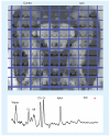
As the concept of a network of injury has emerged in the treatment of epilepsy, the importance of evaluating that network noninvasively has also grown. Recently, studies utilizing magnetic resonance spectroscopic imaging, manganese-enhanced MRI and functional (f)MRI measures of resting state connectivity have demonstrated their ability to detect injury and dysfunction in cerebral networks involved in the propagation of seizures. The ability to noninvasively detect neuronal injury and dysfunction throughout cerebral networks should improve surgical planning, provide guidance for placement of devices that target network propagation and provide insights into the mechanisms of recurrence following resective surgery.
Figures


Similar articles
-
The Interictal Suppression Hypothesis in focal epilepsy: network-level supporting evidence.Brain. 2023 Jul 3;146(7):2828-2845. doi: 10.1093/brain/awad016. Brain. 2023. PMID: 36722219 Free PMC article.
-
Network connectivity separate from the hypothesized irritative zone correlates with impaired cognition and higher rates of seizure recurrence.Epilepsy Behav. 2019 Dec;101(Pt A):106585. doi: 10.1016/j.yebeh.2019.106585. Epub 2019 Nov 4. Epilepsy Behav. 2019. PMID: 31698262
-
Resting-state functional magnetic resonance imaging for surgical planning in pediatric patients: a preliminary experience.J Neurosurg Pediatr. 2017 Dec;20(6):583-590. doi: 10.3171/2017.6.PEDS1711. Epub 2017 Sep 29. J Neurosurg Pediatr. 2017. PMID: 28960172 Free PMC article.
-
Presurgical Mapping of the Language Network Using Resting-state Functional Connectivity.Top Magn Reson Imaging. 2016 Feb;25(1):19-24. doi: 10.1097/RMR.0000000000000073. Top Magn Reson Imaging. 2016. PMID: 26848557 Free PMC article. Review.
-
Neuroimaging of Epilepsy.Continuum (Minneap Minn). 2022 Apr 1;28(2):306-338. doi: 10.1212/CON.0000000000001080. Continuum (Minneap Minn). 2022. PMID: 35393961 Review.
References
-
- Margerison JH, Corsellis JAN. Epilepsy and the temporal lobes. Brain. 1968;91:499–531. - PubMed
-
- Blumenfeld H, McNally KA, Vanderhill SD, et al. Positive and negative network correlations in temporal lobe epilepsy. Cereb. Cortex. 2004;14(8):892–902. - PubMed
-
- Labate A, Cerasa A, Gambardella A, Aguglia U, Quattrone A. Hippocampal and thalamic atrophy in mild temporal lobe epilepsy: a VBM study. Neurology. 2008;71(14):1094–1101. - PubMed
-
- Riederer F, Lanzenberger R, Kaya M, et al. Network atrophy in temporal lobe epilepsy: a voxel-based morphometry study. Neurology. 2008;71(6):419–425. - PubMed
-
- Gong G, Concha L, Beaulieu C, Gross DW. Thalamic diffusion and volumetry in temporal lobe epilepsy with and without mesial temporal sclerosis. Epilepsy Res. 2008;80(2–3):184–193. - PubMed
Grants and funding
LinkOut - more resources
Full Text Sources
Research Materials
