. 2009 Apr;2(2):118-26.
doi: 10.1111/j.1752-8062.2009.00091.x.

Induced pluripotent reprogramming from promiscuous human stemness related factors

Affiliations

Induced pluripotent reprogramming from promiscuous human stemness related factors

Timothy J Nelson et al. Clin Transl Sci. 2009 Apr.

Abstract

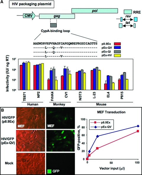
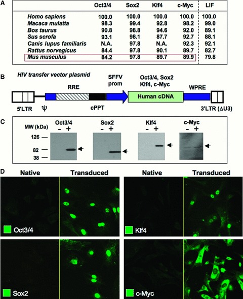
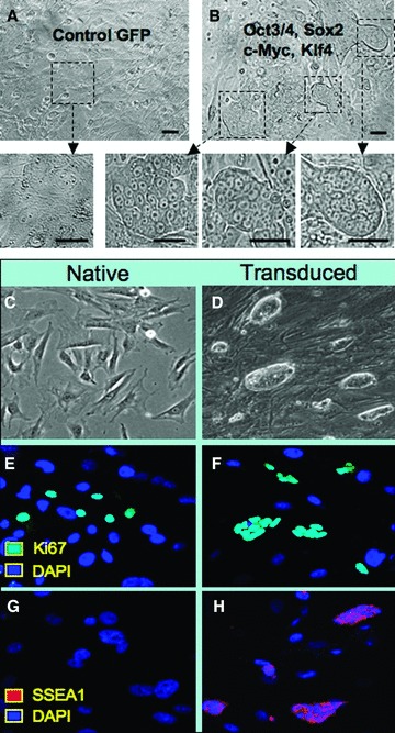
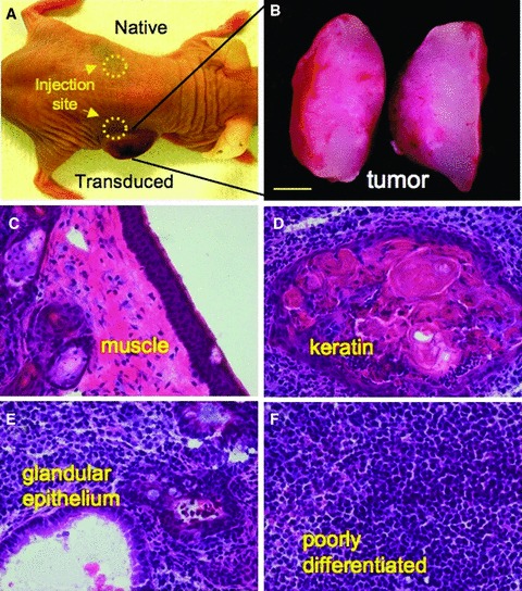
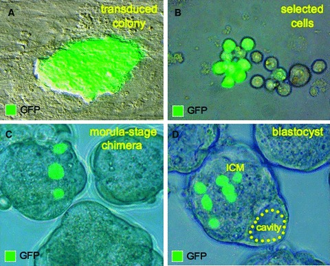
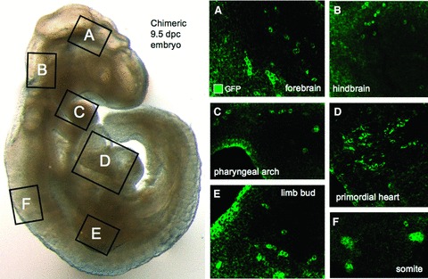
Ectopic expression of pluripotency gene sets provokes nuclear reprogramming in permissive somatic tissue environments generating induced pluripotent stem (iPS) cells. The evolutionary conserved function of stemness orthologs was here tested through interspecies transduction. A spectrum of HIV-based lentiviral vectors was designed, and point mutations in the HIV-1 capsid region identified for efficient infectivity and expanded trans-species tropism. Human pluripotent gene sequences, OCT3/4, SOX2, KLF4 and c-MYC, packaged into engineered lentiviral expression vectors achieved consistent expression in non-human fibroblasts. Despite variation in primary amino-acid sequence between species, introduction of human pluripotent genes produced cell lines with embryonic stem cell-like morphology. Transduced fibroblasts differentiated in vitro into all three germ layers according to gastrulation gene expression profiles, and formed in vivo teratoma with multi-lineage potential. Reprogrammed progeny incorporated into non-human morula to produce blastomeres capable of developing into chimeric embryos with competent organogenesis. This model system establishes a prototypic approach to examine consequences of human stemness factors induced reprogramming in the context of normal embryonic development, exploiting non-human early stage embryos. Thus, ectopic xeno-transduction across species unmasks the promiscuous nature of stemness induction, suggesting evolutionary selection of core processes for somatic tissue reprogramming.

Keywords: HIV; KLF4; OCT3/4; SOX2; c-MYC; chimera; iPS; lentiviral; ortholog.


Figures

Figure 1
Figure 1
The H87Q capsid substitution in vector packaging constructs increases HIV vector infectivity across species. (A) Naturally occurring capsid substitutions were introduced into the cyclophilin A‐binding region of HIV‐1 gag gene of a vector packaging construct, p8.9Ex. Infectious HIV vectors were generated by packaging a GFP‐carrying HIV vector genome with the modified constructs, and the amounts of vectors were normalized by the levels of endogenous reverse transcriptase (RT) activity in vector particles. Human, simian, and murine cell lines were infected with various amounts of GFP‐expressing vectors, and GFP‐positive cell populations were analyzed by flow cytometry. Vector infectivity in each target cell line was determined by infectious units per nanogram RT activity. (B) GFP‐carrying HIV vectors were generated with a conventional HIV packaging construct (p8.9Ex) or a packaging construct with the H87Q capsid subsitution (pEx‐QV). MEFs (5 × 10⁴) were infected with increasing amounts of unconcentrated vectors. The percentage of transfected cells was observed by comparing total cells to GFP‐positive cells under UV microscope 3 days after infection (left panels, with 20 μL of vector input) and analyzed by flow cytometry 5 days after vector infection (right panel).
Figure 2
Figure 2
Efficient expression of sternness‐associated factors in human and murine cell types. (A) Percent amino acid homology among orthologous sternness‐related factors. Homology for LIF served as benchmark. Homology was determined by LALIGN program (EMBNet). NA = protein sequence not available. (B) Scheme of the HIV‐1 vector genome construct used to generate sternness factor‐expressing vectors. Ψ= packaging signal; LTR = long terminal repeat; RRE = Rev‐responsive element; cPPT = central polypurine tract; SFFV = spleen focus‐forming virus promoter; WPRE = Woodchuck hepatitis virus posttranscriptional regulatory element. OCT‐314, SOX2, KLF4, and c‐MYC cDNAs were driven by an internal SFFV promoter. The KLF4‐encoding vector lacks WPRE. (C) 29 3T cells (2 × 10⁵) were infected with 50 μL of the sternness factor‐expressing vectors. Three days after infection, expression of full‐length sternness factors was verified by Western blotting with respective antibodies. (D) MEFs (5 × 10⁴) were infected with 100 μL of unconcentrated vectors. Expression levels of transgene products were visualized by immunostaining 4 days following infection.
Figure 3
Figure 3
Transduced murine fibroblasts with human sternness factors reactivate stem cell phenotype. (A) Control virus expressing GFP demonstrated no discernible changes in MEF morphology. (B) Coinfection with human OCT‐314, SOX2, KLF4, and c‐MYC vectors produced multiple colonies with distinct stem cell‐like morphology that allowed isolation of individual clones. (C) Native MEFs continued to grow in monolayer and displayed contact inhibition at confluence. (D) Clonal expansion of transduced cells demonstrated morphology similar to embryonic stem cells. (E, F) Both native and transduced MEFs expressed markers of cell cycle activation indicated by Ki67 (cyan) in a subpopulation of progeny. (C, H) The stem cell marker SSEA1 (red) was uniquely expressed within transduced cells compared with native MEFs identified by nuclear staining with DAPI (blue).
Figure 4
Figure 4
Gene expression following in vitro differentiation recapitulates gastrulation. (A) Differentiation of trans duced cells was facilitated by three‐dimensional clustering in a hanging drop to allow spontaneous maturation over a 5‐day time course. (B) Pluripotency markers, OCT4, SOX2, and FGF4, were highest in transduced cells at day 0 of differentiation, compared with either native MEFs or transduced counterparts at day 5 post initiation of differentiation. (C) The markers of mesoderm (Gsc), endoderm (Soxl7), and ectoderm (Zicl) were higher after 5 days of differentiation compared with day 0 of differentiation.
Figure 5
Figure 5
Multilineage in vivo differentation within tumors. (A) Spontaneous in vivo differentiation was monitored in immunodeficient mice following subcutaneous injection by comparing native and transduced MEFs. (B) Tumor growth was detected only from sites injected with transduced cells ater 1–2 weeks, followed by rapid expansion of tumor bulk, absent from native MEF injection sites. (C–F) Tissue was harvested at 4–6 weeks post injection. Cryosections and tissue staining demonstrated multiple lineages within the complex architecture of the nascent tumor and included muscle, keratin, glandular epithelium, and poorly differentiated tissues.
Figure 6
Figure 6
Transduced cells integrate into host morula. (A, B) Transduced MEFs were labeled with GFP tag for tracking and allowed selection for ex utero integration into early‐stage embryos. (C) Diploid aggregation between labeled transduced cells and normal morula produced chimeric early embryos. (D) Chimeric embryos developed into blastocysts, which displayed proper cavitation and formation of mosaic inner cell mass (ICM) with GFP‐labeled blastomeres.
Figure 7
Figure 7
Organogenesis derived from transduced cells. (A‐F) Chimeric embryos were transferred into a surrogate mother for in utero differentiation and were harvested at 9.5 dpc for tissue analysis. Confocal microscopy revealed transduced progeny throughout the embryo including neuronal tissues of the forebrain (A) and hindbrain (B), along with the multilineage phaiyngeal arches that contained endoderm derivatives (C). Mesoderm‐derived lineages were present in the heart (D), limb bud (E), and somites (F).

Similar articles

Cited by

References

    1. Wilmut I, Schnieke AE, McWhir J, Kind AJ, Campbell KH. Viable offspring derived from fetal and adult mammalian cells. Nature. 1997; 385(6691): 810–813. - PubMed
    1. Campbell KH, McWhir J, Ritchie WA, Wilmut I. Sheep cloned by nuclear transfer from a cultured cell line. Nature. 1996; 380(6569): 64–66. - PubMed
    1. Hochedlinger K, Jaenisch R. Nuclear reprogramming and pluripotency. Nature. 2006; 441(7097): 1061–1067. - PubMed
    1. Armstrong L, Lako M, Dean W, Stojkovic M. Epigenetic modification is central to genome reprogramming in somatic cell nuclear transfer. Stem Cells. 2006; 24(4): 805–814. - PubMed
    1. Byrne JA, Pedersen DA, Clepper LL, Nelson M, Sanger WG, Gokhale S, Wolf DP, Mitalipov SM. Producing primate embryonic stem cells by somatic cell nuclear transfer. Nature. 2007; 450(7169): 497–502. - PubMed

Publication types

MeSH terms

LinkOut - more resources