. 2009 May 1;4(3):277-293.
doi: 10.2217/fvl.09.7.

Helicase inhibitors as specifically targeted antiviral therapy for hepatitis C

Affiliations

Helicase inhibitors as specifically targeted antiviral therapy for hepatitis C

Craig A Belon et al. Future Virol. .

Abstract

The hepatitis C virus (HCV) leads to chronic liver disease and affects more than 2% of the world's population. Complications of the disease include fibrosis, cirrhosis and hepatocellular carcinoma. Current therapy for chronic HCV infection, a combination of ribavirin and pegylated IFN-alpha, is expensive, causes profound side effects and is only moderately effective against several common HCV strains. Specifically targeted antiviral therapy for hepatitis C (STAT-C) will probably supplement or replace present therapies. Leading compounds for STAT-C target the HCV nonstructural (NS)5B polymerase and NS3 protease, however, owing to the constant threat of viral resistance, other targets must be continually developed. One such underdeveloped target is the helicase domain of the HCV NS3 protein. The HCV helicase uses energy derived from ATP hydrolysis to separate based-paired RNA or DNA. This article discusses unique features of the HCV helicase, recently discovered compounds that inhibit HCV helicase catalyzed reactions and HCV cellular replication, and new methods to monitor helicase action in a high-throughput format.


Figures
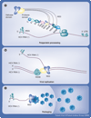
Figure 1
Figure 1. Possible roles of HCV NS3 in viral replication
(A) Polyprotein processing. The protease domain of the NS3 protein is depicted cleaving the viral polyprotein to release mature HCV nonstructural proteins. The helicase could assist this process by interacting with the RNA being translated. (B) RNA replication. The helicase is shown aiding the action of the NS5B polymerase by separating double-stranded RNA duplexes resulting from genome replication. (C) Packaging. The helicase is depicted packaging HCV RNA into virions with the help of the HCV core protein. HCV: Hepaptits C virus; IRES: Internal ribosome entry site; NS: Nonstructural.
Figure 2
Figure 2. Ligand-binding clefts and key amino acids in the HCV NS3 helicase
(A) Model of HCV helicase unwinding DNA. The full-length NS3 structure (Protein Data Bank [PDB] accession number 1CU1) was aligned with the costructure of a related helicase–DNA complex (archaeal Hel308, PDB file 2P6R) to reveal how NS3 might split a duplex. Shown are the NS3 protease (green), NS4A (blue), helicase domain 1 (yellow), domain 2 (purple) and domain 3 (tan), along with the DNA (sticks) bound to Hel308, which is not shown. (B) Putative ATP-binding site. An alignment of the crystal structure of HCV NS3h (PDB file 1A1V) with that of a related helicase cocrystallized with a nonhydrolyzable ATP analog (Dengue virus NS3, PDB 2JLV) to show the coordination of ATP. K210 is in SF2 helicase motif I and coordinates the γ-phosphate of ATP. D290 is in SF2 motif II and coordinates the bridging divalent metal cation (Mg2+). R467 is a likely ‘arginine finger’ that might coordinate the γ-phosphate in the transition state and E291 is the catalytic base that activates the water that attacks the γ-phosphate. The ATP analog is depicted as sticks. (C) Known RNA-binding site. The nucleic acid-binding cleft of PDB file 1A1V is shown as an electrostatic surface (red, negatively charged; blue, positively charged). The surface was calculated without DNA bound, and the DNA was added back to mark the binding site. The oligonuclotide is held in a positively charged pocket centered on E493, held in place by the Arg-clamp motif (R393). (D) Phe-loop motif. A β-loop (ribbon) containing the conserved Phe-loop motif acts to separate double-stranded nucleic acids. (E) Possible second RNA-binding site in the cleft between the protease and helicase domain 2. The surface is colored by domains as in (A). Molecular models were aligned using UCSF Chimera [201], ligands were docked using UCSF DOCK [202], electrostatic surface was calculated with APBS [203] and models were rendered using pyMol [204]. APBS: Adaptive Poisson-Botlzmann Solver; HCV: Hepatits C virus; NS: Nonstructural; SF: Superfamily; UCSF: University of California, San Francisco.
Figure 3
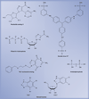
Figure 3. Compounds that likely bind to the ATP site
DRBT: 5,6-dichloro-1-(β-d-ribofuranosyl)benzotriazole; TBBT: 4,5,6,7-tetrabromobenzotriazole. Data taken from [–110].
Figure 4
Figure 4. Compounds that likely to bind the RNA site
(A) QU663 [111]. (B) The p14 peptide RRGRTGRGRRGIYR [59].
Figure 5
Figure 5. Compounds binding to unknown sites
(A) DBMTr (3,7-dibromo-5-morpholinomethyltropolone) [113]. (B) Acridone-4-carboxylic acid derivative: 27,9-oxo-9,10-dihydro-acridine-4-carboxylic acid pyridin-2-ylamine. (C) Acridone-4-carboxylic acid derivative: 20, 9-oxo-9,10-dihydro-acridine-4-carboxylic acid pyridin-4-ylamine [115].

Similar articles

Cited by

References

Bibliography

    1. HCV Working Group. Global burden of disease (GBD) for hepatitis C. J. Clin. Pharmacol. 2004;44:20–29. - PubMed
    1. McHutchison JG. Understanding hepatitis C. Am. J. Manag. Care. 2004;10:S21–S29. - PubMed
    1. Habib M, Mohamed MK, Abdel-Aziz F, et al. Hepatitis C virus infection in a community in the Nile delta: risk factors for seropositivity. Hepatology. 2001;33:248–253. - PubMed
    1. Maheshwari A, Ray S, Thuluvath PJ. Acute hepatitis C. Lancet. 2008;372:321–332. - PubMed
    1. Hu KQ, Tong MJ. The long-term outcomes of patients with compensated hepatitis C virus-related cirrhosis and history of parenteral exposure in the United States. Hepatology. 1999;29:1311–1316. - PubMed

Websites

    1. UCSF Chimera: an extensive molecular modeling system. www.cgl.ucsf.edu/chimera/
    1. UCSF DOCK. http://dock.compbio.ucsf.edu/
    1. Adaptive Poisson-Boltzmann solver. http://apbs.sourceforge.net/
    1. PyMOL. www.pymol.org/

LinkOut - more resources