. 2009 Dec 1;14(6):331-340.
doi: 10.1016/j.jala.2009.06.003.

Perspectives on utilizing unique features of microfluidics technology for particle and cell sorting

Affiliations

Perspectives on utilizing unique features of microfluidics technology for particle and cell sorting

Jonathan D Adams et al. JALA Charlottesv Va. .

Abstract

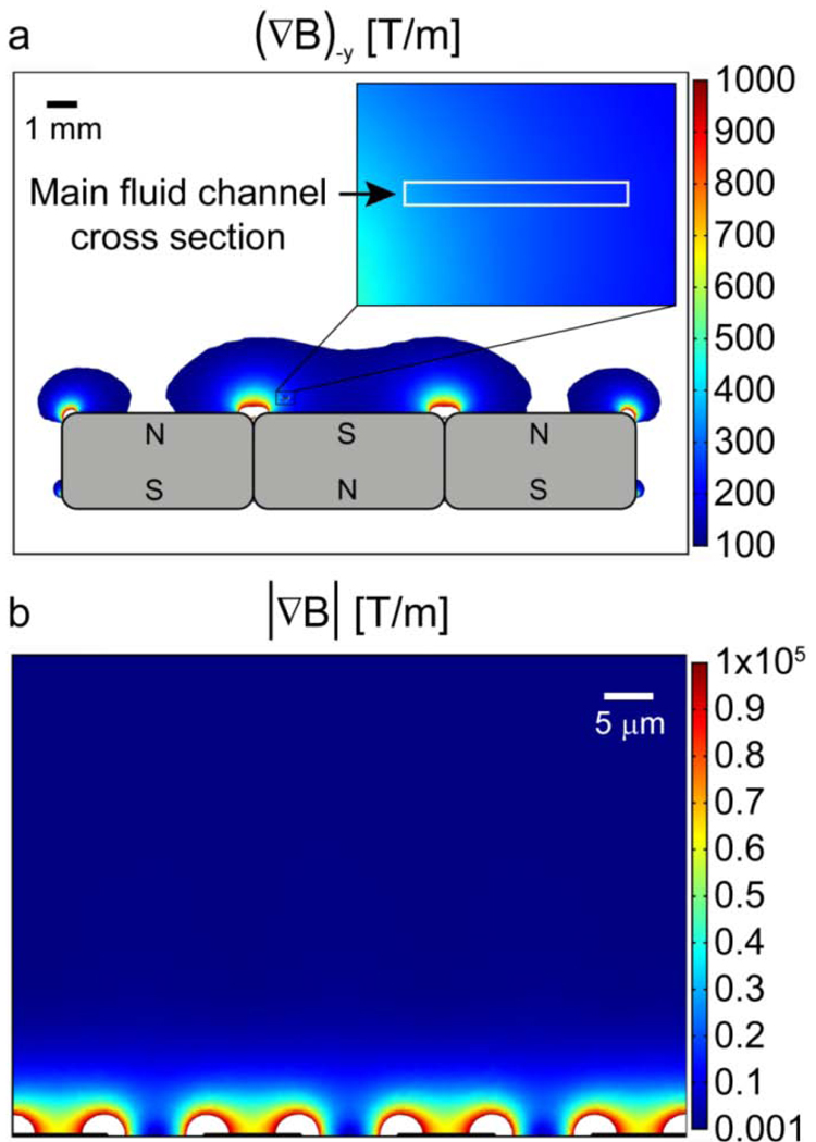
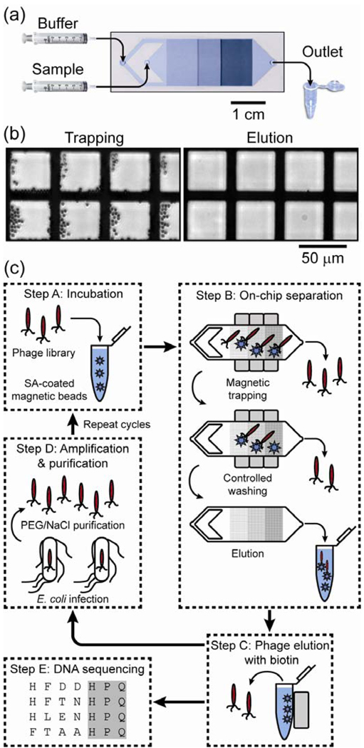
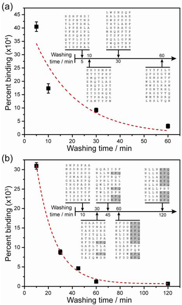
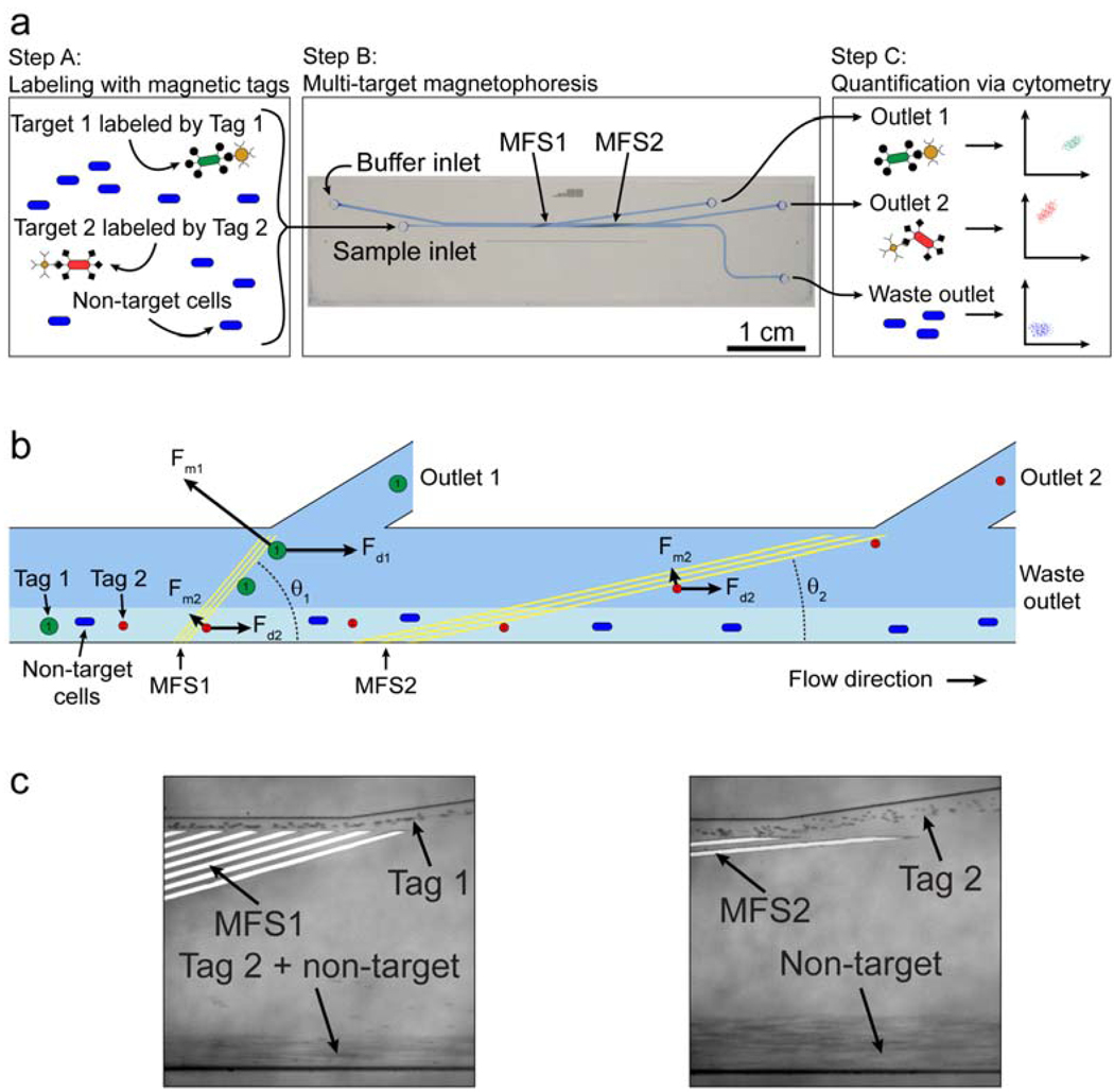
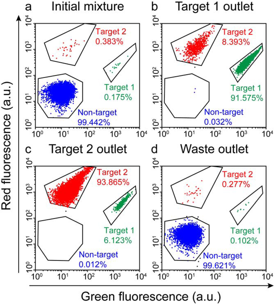
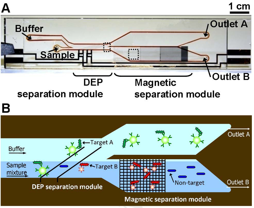
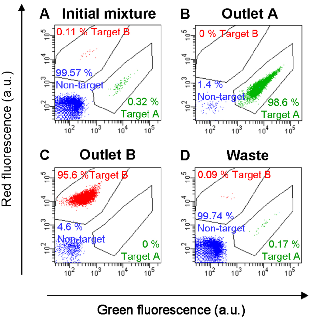
Sample preparation is often the most tedious and demanding step in an assay, but it also plays an essential role in determining the quality of results. As biological questions and analytical methods become increasingly sophisticated, there is a rapidly growing need for systems that can reliably and reproducibly separate cells and particles with high purity, throughput and recovery. Microfluidics technology represents a compelling approach in this regard, allowing precise control of separation forces for high performance separation in inexpensive, or even disposable, devices. In addition, microfluidics technology enables the fabrication of arrayed and integrated systems that operate either in parallel or in tandem, in a capacity that would be difficult to achieve in macro-scale systems. In this report, we use recent examples from our work to illustrate the potential of microfluidic cell- and particle-sorting devices. We demonstrate the potential of chip-based high-gradient magnetophoresis that enable high-purity separation through reversible trapping of target particles paired with high-stringency washing with minimal loss. We also describe our work in the development of devices that perform simultaneous multi-target sorting, either through precise control of magnetic and fluidic forces or through the integration of multiple actuation forces into a single monolithic device. We believe that such devices may serve as a powerful "front-end" module of highly integrated analytical platforms capable of providing actionable diagnostic information directly from crude, unprocessed samples - the success of such systems may hold the key to advancing point-of-care diagnostics and personalized medicine.


Figures

Figure 1
Figure 1
Numerical simulation of long-range and short-range magnetic field gradients in a microfluidic device designed for magnetophoretic separation. a) Long-range gradients are produced by a series of external permanent magnets, with the magnitude of the -y direction gradient exceeding 200 T/m over the cross-section of a microfluidic channel located 0.5 mm above the surface of the magnets. b) An abrupt change in relative permittivity (μr) between microfabricated nickel features (μr ~200) and the biological sample (μr ~1) creates an extremely large short-range magnetic field gradient. The magnitude of this gradient is > 104 T/m within 8 μm of the microfabricated features. Image adapted from Adams et al. with permission. ©PNAS 2008.
Figure 2
Figure 2
Overview of the Micro-Magnetic Separation (MMS) device and its application for high-purity phage library screening. a) Micrograph of the MMS device showing the channel design, nickel pattern and flow path. The device dimensions are 64 mm ×15.7 mm × 1.5 mm (L × W × H), and the height and width of the microfluidic channel are 30 μm and 12 mm, respectively. b) Bright field optical micrographs of the nickel pattern in the microchannel. When an external field is applied (left), the large magnetic field gradients at the edges of the nickel pattern effectively trap the beads, but when the external field is removed (right), the nickel pattern is de-magnetized and the beads are efficiently eluted. c) Selection of the phage display library using the MMS device. Step A: The phage library is mixed and incubated with target molecule-conjugated magnetic beads. Step B: NeFeB permanent magnets are applied to the MMS device to specifically trap phage particles bound to the magnetic beads, which are subsequently held in place and washed under controlled conditions to eliminate nonspecific interactions. The nickel patterns are then de-magnetized, and the phage-carrying beads are eluted. Step C: The phage are dissociated from the protein-conjugated beads by competitive elution with biotin. Step D: Isolated phage are amplified via infection of E. coli cells, and subsequently purified with PEG/NaCl solution for additional rounds of selection or analysis. Step E: Clones from each round of selection are randomly picked and their DNA is sequenced. Figure reprinted from Liu et al. with permission of the authors.
Figure 3
Figure 3
The importance of controlling the washing conditions during phage selection. a) In the first round of selection, the percentage of recovered phage as a function of washing time decays non-linearly, as non-specifically bound and weak binding phage are removed. When modeled as a first order exponential (dashed line), the dissociation rate constant was kd1 = 1.0±0.1∙10−3 s−1. (Inset) The canonical target-binding peptide motif (HPQ) was not found in clones isolated after the first round. b) In the second round, the percentage of bound phage also showed an exponential decay as stringency (washing time) increased, with a remarkably similar dissociation rate constant of kd2 = 1.07±0.04∙10−3 s−1. (Inset) The percentage of clones with the HPQ motif increased monotonically as a function of washing time; after 120 minutes of washing, 8 out of 9 clones contained this motif. Figure reprinted from Liu et al. with permission of the authors.
Figure 4
Figure 4
MT-MACS separation architecture. a) The separation process. Step A: The sample contains an excess of non-target cells and two different target cell types, which are labeled with two different magnetic tags via specific surface markers. Step B: The sample is continuously pumped into the device, where the two target cell types are sorted into spatially segregated, independent outlets at regions of high magnetic field gradient generated by two sets of microfabricated ferromagnetic strips (MFS1 and MFS2). Step C: The eluted fractions from each outlet are analyzed via flow cytometry. b) A free-body diagram showing the balance of forces at the MFS structures. At MFS 1 (θ1 =15°), tag 1-labeled cells are deflected and elute through outlet 1 because Fm1>Fd1sin(θ1) . This is not the case for tag 2-labeled target 2 cells, which are instead deflected at MFS 2 (θ2 =5°), where Fm2 >Fd2 sin(θ2) and elute through outlet 2. Non-target cells are not deflected by either MFS and elute through the waste outlet. c) Optical micrographs (100X magnification) of tags being separated at the two MFS structures at a total flow rate of 47 ml/hr (sample = 5 ml/hr, buffer = 42 ml/hr). Tag 1 is deflected at MFS 1 (left), while tag 2 is deflected by MFS2 (right). Figure taken from Adams et al. with permission of the authors. © PNAS 2008
Figure 5
Figure 5
Cytometric analysis of simultaneous,high-purity enrichment of multiple bacterial target cell types in the MT-MACS device. a) The initial sample mixture consists of 99.442% non-target cells (expressing BFP) doped with 0.175% target 1 cells and 0.383% target 2 cells. b) The cell mixture recovered at the target 1 outlet consisted of 91.575% target 1 cells, 8.393% target 2 cells, and 0.032% non-target cells. c) The output at the target 2 outlet was comprised of 93.865% target 2 cells, 6.123% target 1 cells, and 0.012% non-target cells. d) Waste outlet output consisted of 99.621% non-target cells, 0.102% target 1 cells and 0.277% target 2 cells. Figure taken from Adams et al. with permission of the authors. © PNAS 2008
Figure 6
Figure 6
Multi-target bacterial cell sorting via iDMACS. a) Photograph of the fabricated idMACS device. Overall device size is 7 cm × 1.5 cm, including both DEP and magnetic separation modules. b) The physics of multi-target separation via iDMACS. Target A cells, labeled with DEP tags, are deflected at a set of angled electrodes to elute through outlet A. Subsequently, an array of ferromagnetic strips captures magnetically-tagged target B cells, which are then eluted through outlet B after washing. Unlabeled non-target cells are neither deflected by the electrodes nor captured by the strips. Figure reprinted from Kim et al. with permission of the authors.
Figure 7
Figure 7
Multi-target bacterial cell sorting performance using the iDMACS device. a) Two-color flow cytometry measurement of the initial sample, which consisted of an excess of non-target cells (99.57%) with low concentrations of labeled target A (0.32%) and target B (0.11% ) cells. b) After a single round of separation, the outlet A fraction contained almost exclusively target A cells (98.6%, a 310-fold enrichment), and no target B cells (0%). c) Conversely, the outlet B fraction contained primarily target B cells (95.6%, a 870-fold enrichment) and no target A cells (0%). d) The fraction collected at the waste outlet consisted of small quantities of target A (0.17%) and target B (0.09%) cells and mostly non-target cells (99.74%). Figure reprinted from Kim et al. with permission of the authors.

References

    1. West J, Becker M, Tombrink S, Manz A. Micro total analysis systems: latest aachievements. Anal. Chem. 2008;80(12):4403–4419. - PubMed
    1. Lee HJ, Goodrich TT, Corn RM. SPR imaging measurements of 1-D and 2-D DNA microarrays created from microfluidic channels on gold thin films. Anal. Chem. 2001;73(22):5525–5531. - PubMed
    1. Margu1ies M, Egholm M, Altman WE, Attiya S, Bader JS, Bemben LA, Berka J, Braverman MS, Chen YJ, Chen ZT, Dewell SB, Du L, Fierro JM, Gomes KV, Godwin BC, He W, Helgesen S, Ho CH, Irzyk GP, Jando SC, Alenquer MLI, Jarvie TP, Jirage KB, Kim JB, Knight JR, Lanza JR, Leamon JH, Lefkowitz SM, Lei M, Li J, Lohman KL, Lu H, Makhijani VB, McDade KE, McKenna MP, Myers EW, Nickerson E, Nobile JR, Plant R, Puc BP, Ronan MT, Roth GT, Sarkis GJ, Simons JF, Simpson JW, Srinivasan M, Tartaro KR, Tomasz A, Vogt KA, Volkmer GA, Wang SH, Wang Y, Weiner MP, Yu PG, Begley RF, Rothberg JM. Genome sequencing in microfabricated high-density picolitre reactors. Nature. 2005;437(7057):376–380. - PMC - PubMed
    1. Yager P, Edwards T, Fu E, Helton K, Nelson K, Tam MR, Weigl BH. Microfluidic diagnostic technologies for global public health. Nature. 2006;442(7101):412–418. - PubMed
    1. Hansel TT, Devries IJM, Iff T, Rihs S, Wandzilak M, Betz S, Blaser K, Walker C. An Improved Immunomagnetic Procedure for the isolation of highly purified human blood eosinophils. J. Immunol. Meth. 1991;145(1–2):105–110. - PubMed

LinkOut - more resources