

Glycoprofiling of the Human Salivary Proteome
- PMID: 20161393
- PMCID: PMC2782851
- DOI: 10.1007/s12014-008-9021-0
Glycoprofiling of the Human Salivary Proteome
Abstract
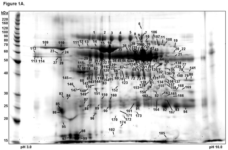
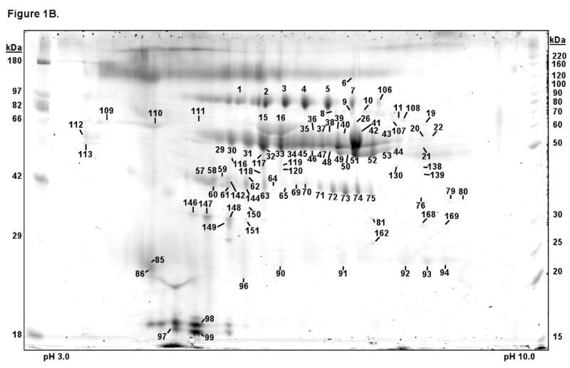
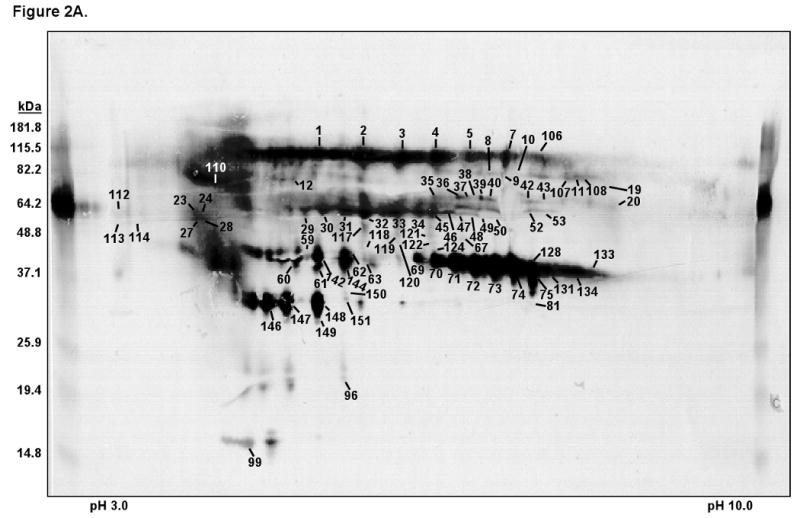
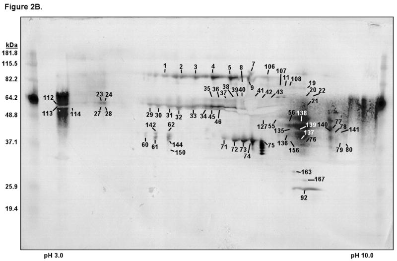
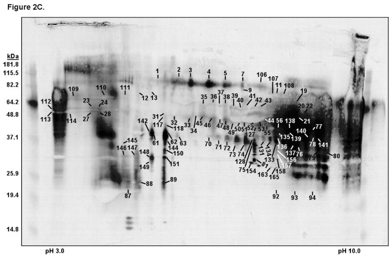
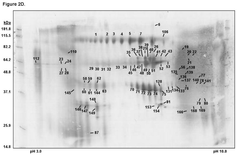
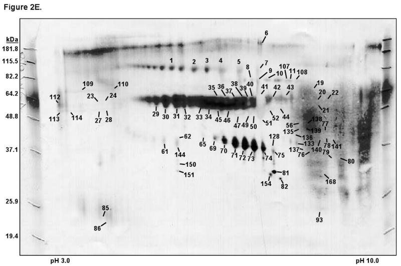
Glycosylation is important for a number of biological processes and is perhaps the most abundant and complicated of the known post-translational modifications found on proteins. This work combines two-dimensional polyacrylamide gel electrophoresis (2-DE) and lectin blotting to map the salivary glycome, and mass spectrometry to identity the proteins that are associated with the glycome map. A panel of 15 lectins that recognize six sugar-specific categories was used to visualize the type and extent of glycosylation in saliva from two healthy male individuals. Lectin blots were compared to 2-D gels stained either with Sypro Ruby (protein stain) or Pro-Q Emerald 488 (glycoprotein stain). Each lectin shows a distinct pattern, even those belonging to the same sugar-specific category. In addition, the glycosylation profiles generated from the lectin blots show that most of the salivary proteins are glycosylated and that the pattern is more widespread than is demonstrated by the glycoprotein stained gel. Finally, the co-reactivity between two lectins was measured to determine the glycan structures that are most and least often associated with one another along with the population variation of the lectin reactivity for 66 individuals.
Figures
Similar articles
-
Functional characterization of two-dimensional gel-separated proteins using sequential staining.Electrophoresis. 2005 Jan;26(1):225-37. doi: 10.1002/elps.200406176. Electrophoresis. 2005. PMID: 15624177
-
Rapid and simple single nanogram detection of glycoproteins in polyacrylamide gels and on electroblots.Proteomics. 2001 Jul;1(7):841-55. doi: 10.1002/1615-9861(200107)1:7<841::AID-PROT841>3.0.CO;2-E. Proteomics. 2001. PMID: 11503209
-
Enrichment and identification of glycoproteins in human saliva using lectin magnetic bead arrays.Anal Biochem. 2016 Mar 15;497:76-82. doi: 10.1016/j.ab.2015.11.024. Epub 2015 Dec 30. Anal Biochem. 2016. PMID: 26743719
-
Glycosylation of secretory proteins in salivary glands and saliva studied by lectin-probes.Eur J Morphol. 1996 Aug;34(3):177-80. doi: 10.1076/ejom.34.3.177.13034. Eur J Morphol. 1996. PMID: 8874092 Review.
-
Lectin Histochemistry: Historical Perspectives, State of the Art, and the Future.Methods Mol Biol. 2017;1560:93-107. doi: 10.1007/978-1-4939-6788-9_6. Methods Mol Biol. 2017. PMID: 28155147 Review.
Cited by
-
The scientific exploration of saliva in the post-proteomic era: from database back to basic function.Expert Rev Proteomics. 2012;9(1):85-96. doi: 10.1586/epr.11.80. Expert Rev Proteomics. 2012. PMID: 22292826 Free PMC article. Review.
-
Characterization of the human submandibular/sublingual saliva glycoproteome using lectin affinity chromatography coupled to multidimensional protein identification technology.J Proteome Res. 2011 Nov 4;10(11):5031-46. doi: 10.1021/pr200505t. Epub 2011 Oct 13. J Proteome Res. 2011. PMID: 21936497 Free PMC article.
-
Protein analysis by shotgun/bottom-up proteomics.Chem Rev. 2013 Apr 10;113(4):2343-94. doi: 10.1021/cr3003533. Epub 2013 Feb 26. Chem Rev. 2013. PMID: 23438204 Free PMC article. Review. No abstract available.
-
Salivary Protein Roles in Oral Health and as Predictors of Caries Risk.Open Life Sci. 2018 May 18;13:174-200. doi: 10.1515/biol-2018-0023. eCollection 2018 Jan. Open Life Sci. 2018. PMID: 33817083 Free PMC article.
-
Effect of mechanically stimulated saliva on initial human dental biofilm formation.Sci Rep. 2019 Aug 14;9(1):11805. doi: 10.1038/s41598-019-48211-3. Sci Rep. 2019. PMID: 31413280 Free PMC article.
References
-
- Slavkin HC. Toward molecularly based diagnostics for the oral cavity. J Am Dent Assoc. 1998;129:1138–1143. - PubMed
-
- Oppenheim FG, Salih E, Siqueira WL, Zhang W, Helmerhorst EJ. The Salivary Proteome and Its Genetic Polymorphisms. Ann NY Acad Sci. 2007;1098:22–50. - PubMed
-
- Guo T, Rudnick PA, Wang W, Lee CS, et al. Characterization of the human salivary proteome by capillary isoelectric focusing/nanoreversed-phase liquid chromatography coupled with ESI-tandem MS. J Proteome Res. 2006;5:1469–1478. - PubMed
-
- Hirtz C, Chevalier F, Centeno D, Egea JC, et al. Complexity of the human whole saliva proteome. J Physiol Biochem. 2005;61:469–480. - PubMed
-
- Walz A, Stuhler K, Wattenberg A, Hawranke E, et al. Proteome analysis of glandular parotid and submandibular-sublingual saliva in comparison to whole human saliva by two-dimensional gel electrophoresis. Proteomics. 2006;6:1631–1639. - PubMed
Grants and funding
LinkOut - more resources
Full Text Sources
Other Literature Sources