

doi: 10.1016/j.pnmrs.2009.07.002.
Recent Advances in the Application of Solution NMR Spectroscopy to Multi-Span Integral Membrane Proteins
Affiliations
- PMID: 20161395
- PMCID: PMC2782866
- DOI: 10.1016/j.pnmrs.2009.07.002
Item in Clipboard
Recent Advances in the Application of Solution NMR Spectroscopy to Multi-Span Integral Membrane Proteins
Prog Nucl Magn Reson Spectrosc.
.
No abstract available
Figures
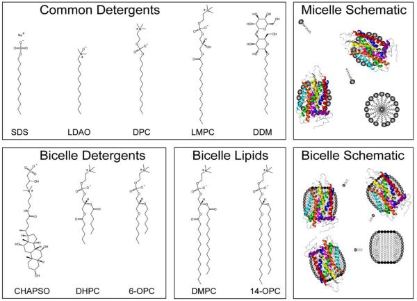
Common detergents and lipids used to prepare micelle and bicelle samples for solution state NMR studies of membrane proteins. Several common micelle forming detergents are shown on the top row (left to right): SDS, LDAO, DPC, LMPC, DDM. Detergents for forming bicelles: CHAPSO, DHPC, 6-OPC. Lipids for forming bicelles: DMPC, 14-OPC. Shown in the right column are schematics of the morphologies for the micelle and bicelle solutions.
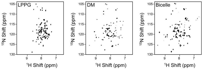
Spectral comparison of 1H-15N TROSY spectra of Smr at 900MHz in LPPG micelles, DM micelles, and q=0.33 DHPC/DMPC isotropic bicelles. Reprinted from Biochimica et Biophysica Acta, 1768, S. F. Poget and M. E. Girvin, “Solution NMR of membrane proteins in bilayer mimics: small is beautiful, but sometimes bigger is better”, 3098-3106, (2007), with permission from Elsevier.
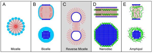
Schematic representation of various membrane mimetics: A. Detergent Micelle B. Ideal Bicelle C. Reverse Micelle D. Nanodisc E. Amphipol. Grey shading designates non-polar environment.
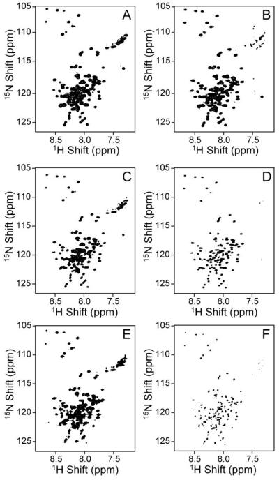
Comparison of 1H-15N HSQC (left panels) and TROSY spectra (right panels) of the uniformly 1H/15N labeled, 99 residue transmembrane C-terminal domain of the amyloid precursor protein (C99) in LMPG micelles (70 kDa complex) acquired at 600MHz (top panels), 800MHz (middle panels), and 900MHz (bottom panels). A. HSQC, 600MHz B. TROSY, 600MHz C. HSQC, 800MHz D. TROSY, 800MHz E. HSQC, 900MHz F. TROSY, 900MHz. Spectra were acquired at 45°C in 5% LMPG, 250mM imidazole, and 1mM EDTA at pH 6.5 [91].
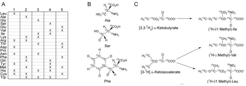
Schematics of several unconventional isotopic labeling schemes used for bacterial expression systems. A. Sample labeling scheme for Combinatorial Selective Labeling (CSL) illustrating the use of five samples used to provide unambiguous discrimination of the amino acid types. B. A selective representation of the amino acids used in Stereo-Array Isotopic Labeling (SAIL) C. Metabolic precursors for the selective methyl-protonation of Ile, Val, and Leu.
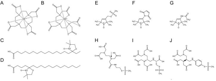
Commonly used paramagnetic probes in solution NMR. Chelated, hydrophilic gadolinium(III) contrast agents: A. Gd-DOTA B. Gd-DTPA. Hydrophobic contrast agents: C. 16-Doxyl-stearic acid (16-DSA) D. 5-DSA. Thiol-reactive nitroxide-based paramagnetic tags: E. 1-Oxyl-2,2,5,5-tetramethylpyrroline-3-methylmethanethiosulfonate (MTSL) F. 1-Oyl-,2,2,5,5-tetramethylpyrrolidine-3-maleimide (3-Maleimido-PROXYL) G. 3-(2-Iodoacetamido)-PROXYL. Thiol-reactive metal chelation tags: H. MTS-EDTA. (Arrow indicates the chiral center which forms upon metal ion complexation) Fixed chirality thiol reactive chelation tags: I. N-[(R)-2,3-Bis[di(carboxymethyl)amino]propionyl]-S-mesyl-(R)-cysteine (R,R-2) J. N,N'-dithiodi(4,1-phenylene)bis-(S)-2,3-bis[di(tert-butoxycarbonylmethyl)amino] propanamide (ent-5). [334]
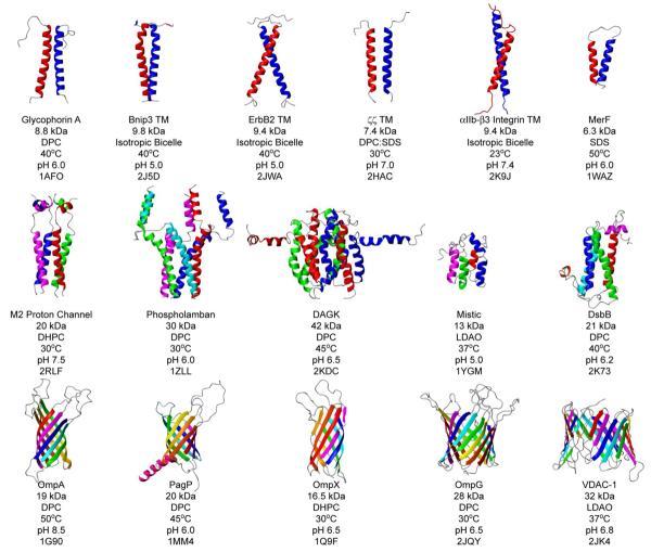
Gallery of structures determined by solution state NMR spectroscopy as of April 2009. Structures are listed with their respective protein mass, the membrane mimetic for which the structure was determined, the temperature used, the pH used, and the deposited PBD ID Number. Ribbon models prepared using MOLMOL [421].
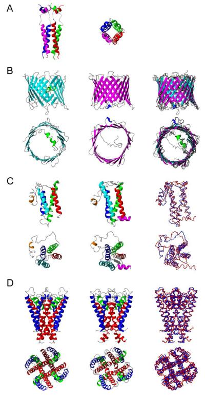
Structures determined by solution state NMR of selected IMPs. A. Tetrameric M2 proton channel of the influenza A virus in DPC micelles. B. Structures determined for the human voltage-dependent anion channel (VDAC-1) in LDAO micelles. Left. VDAC-1 determined from combined NMR/X-ray data (PDB 2JK4). Center. VDAC-1 determined exclusively from NMR data (PDB 2K4T). Right. Overlay of two NMR-derived VDAC-1 structures onto the x-ray structure of mouse VDAC-1 shown in black (PDB 3EMN). C. DsbB structures. Left. DsbB structure determined by x-ray crystallography (PDB 2HI7). Center. DsbB structure determined by NMR in DPC micelles (PDB 2K73). Right. Overlay of the two DsbB structures: PDB 2H17 in blue, PDB 2K73 in red. D. KcsA structures. Left. KcsA structure determined by x-ray (PDB 2K4C). Center. KcsA WSK-3 mutant structure solved by NMR (PDB 2K1E). Right. Overlay of the two KcsA structures: PDB 2K4C in blue, PDB 2K1E in red. Models prepared using MOLMOL [421].
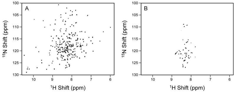
Spectral comparison of a microbial GPCR-like seven transmembrane sensory rhodopsin (pSRII) and a human GPCR. A. 1H,15N-TROSY spectrum of pSRII in DHPC micelles acquired at 600MHz at 50°C and pH 5.9. (from [218]; A. Gautier, J. P. Kirkpatrick, and D. Nietlispach: “Solution-state NMR spectroscopy of a seven-helix transmembrane protein receptor: backbone assignment, secondary structure, and dynamics”. Angewandte Chemie, International Edition in English. (2008), 47, 7297-7300. Copyright Wiley-VCH Verlag GmbH & Co. KGaA. Reproduced with permission. B. 1H-15N TROSY of the human kappa-opioid receptor type 1, 380 residues, acquired at 800MHz in LMPC micelles at 18°C and pH 7.4.
References
-
- Goffeau A, Barrell BG, Bussey H, Davis RW, Dujon B, Feldmann H, Galibert F, Hoheisel JD, Jacq C, Johnston M, Louis EJ, Mewes HW, Murakami Y, Philippsen P, Tettelin H, Oliver SG. Science. 1996;274:546. - PubMed
-
- Jimonet P, Jager R. Curr. Opin. Drug. Discov. Devel. 2004;7:325. - PubMed
-
- Schnur DM, Hermsmeier MA, Tebben AJ. J. Med. Chem. 2006;49:2000. - PubMed
Grants and funding
LinkOut - more resources
Full Text Sources
Other Literature Sources
